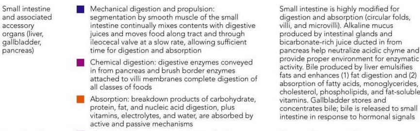
Mostrando entradas con la etiqueta DIGESTIVE SYSTEM. Mostrar todas las entradas

jueves, 9 de abril de 2015

Nausea, vomiting and acute gastrointestinal events

NAUSEA AND VOMITING
Consider the underlying mechanisms and potential causes: 
-establish the history (when and how did it start, other symptoms or illnesses, pain, has it happened before, anyone else got the same symptoms?).
-is this gastrointestinal disease (any diarrhoea, abdominal pain, obstruction, is the cause acute liver damage, alcohol abuse?)
-is it drug-related? (any new medication, digoxin toxicity or opiate side-effect?)
-could this be raised intracranial pressure? (headache, malignant disease or altered consciousness level?)
-could this be metabolic? (renal failure, sodium and calcium levels, ketoacidosis?).

Nausea and vomiting in acute medical conditions:
-Migraine: commonly accompanied by vomiting. Other causes of headache like meningitis, increased intracranial pressure and subarachnoid haemorrhage are also associated with vomiting.
-Myocardial infarction: vomiting is an important feature that distinguishes the pain of a myocardial infarction from that of angina and is often accompanied by nausea and sweating.
-Sepsis: vomiting can be the only symptom of hidden infection, particularly in infections in the kidneys and lower urinary tract.
-Acute gastric dilatation: gross gastric distension leads to upper abdominal swelling associated with nausea, often accompanied by hiccups and belching. A common outcome is sudden vomiting, aspiration an cardiac arrest.


ACUTE UPPER GASTROINTESTINAL HAEMORRHAGE

Causes:
-Helicobacter pylori infection and bleeding peptic ulcers
-Acute gastritis, duodenitis and acute erosions (stress ulcers)
-Mallory-Weiss tear:
-Oesophageal varices
-Gastric and oesophageal cancer
-Dieulafoy´s erosion: erosion of a congenitally abnormal artery in the lining of the stomach, emergecy surgery is often required.

Management:
-Ensuring the safety of the patient: ABCDE.
-Investigations: endoscopy.
-Assessing the degree of bleeding: history (presence of melaena with haematemesis, clots in the vomit and recognisable blood in the stool, bleeding accompanied by syncope, postural dizziness and overwhelming thirst suggest a major bleed) and examination (pale and clammy, tachycardia of more than 100 beats/min , systolic blood pressure of less than 100mmHg and postural hypotension).
-Risk assessment with the Rockall Score:


Important nursing tasks: 
-ensure the safety of the patient, assess the likely severity of the initial blood loss and report any further loss, save any important evidence (vomited blood, fresh melaena), report any abdominal pain.
-correct the oxygen saturation,
-secure the venous access and ensure accurate fluid balance charts,
-complete baseline observations, measure the pulse, blood pressure and respiratory rate. Report any changes on vital signs which should be checked at appropiate intervals.
-identify the signs of shock (pallor, sweating, restlessness, confusion),
-take an appropiate history, ask about liver disease and clotting disorders,
-reassure the patient and attend to patient´s basic comforts, warn about the likely need for an endoscopy.

ACUTE LIVER FAILURE AND HEPATIC ENCEPHALOPATHY
Acute liver failure can be precipitated by an acute event such as viral hepatitis or by a background of chronic liver disease with portal hypertension.

Components of acute liver failure:
-impairment of the consciousness level: the combination of poor liver function and portosystemic shunting means that toxic substances are either dealt with ineffectively by the liver or bypass it altogether. The result is a toxic encephalopathy with imapirment of the consciousness level that can progress rapidly from confusion and agitation to deep coma. It is useful to assess and follow changes using the Hepatic encephalopathy scoring system:


-bleeding varices and clotting abnormalities: patients in acute liver failure are at great risk from upper gastrointestinal bleeding due to the combination of varices and impaired clotting.
-sepsis: infection can trigger or complicate acute liver failure.
-metabolic abnormalities: the main problems are hypoglycaemia and renal failure.

Critical nursing tasks in acute liver failure:
-Ensure the safety of the patient: ABCDE.
-Gain venous access.
-Carry out top-to-toe examination.
-Liaise with patient´s family.
-Ascertain the possible causes of acute liver failure: the most common are paracetamol poisoning and acute hepatitis.
-Explain the likely interventions in the first 48 h: endoscopy, radiology, transfusions, multiple infusion lines and possible transfers.
-Obtain a drug history.

Other important nursing tasks are: determine accurate fluid balance, identify sepsis, reassure the patient and manage confusion and disorientation, carry out general nursing measures.

ACUTE ABDOMINAL PAIN
Abdominal pain is a common complaint on the acute medical ward and it is a feature of several medical conditions.

Nursing tasks:
-Establish the site and pattern of the pain.
-Assess the severity of the pain.
-Look for signs of shock (tachycardia, hypotension and increased respiratory rate).
-Examine for abdominal rigidity and peritonitis: the most obvious features of acute peritonitis are localised pain on coughing and board-like abdominal rigidity, the patient does not want to move, appears shocked and is usually vomiting. Acute peritonitis may be accompanied by signs of sepsis (hypotension, rapid thready pulse and cold, mottled extremities).
-Prepare for resuscitation: the basic requirements are oxygen, IV fluids, nasogastric intubation and urinary catheterisation.

Also, it is important to look at associated clinical features, prepare the patient for further investigations, review the previous history, the urine test results and the stool chart.


ACUTE DIARRHOEA

Infective diarrhoea:


Assessment:
-History of recent foreign travel
-Ask about other possible cases
-Have there been recent doubtful meals/mass catering...?)
-History of recent antibiotic therapy
-Is this patient at special risk?: elderly, diabetic, immunosuppressed, cardiac and kidney disease.
-Is there a history of recurrent diarrhoea?: inflammatory bowel disease.

Nursing assessment: ABCDE, assess fluid loss, look for evidence of shock and diagnosis from stool cultures.

Nursing management: 
-Infection control.
-Correct fluid deficit and keep up with the loss of fluid in the stool and vomit (fluid balance and stool chart).
-Antibiotics are rarely needed unless there are signs of complications: persistent fever with a failure to improve, development of severe colitis or shock, signs of infection, severe colitis with the risk of haemorrhage, toxic dilatation and perforation or HUS (Haemolytic Uraemic Syndrome).

Clostridium Difficile diarrhoea
The indiscriminate use of antibiotics has led to a large rise in bowel infections due to clostridium difficile. The clinical picture varies from mild diarrhoea to severe colitis with fever, systemic sepsis and death.


Diagnosis relies on two fresh stool specimens from any patient with mushy or liquid stools beng sent urgently to the labwith a request for the C.difficile toxin. The result should be available on the same day. Specific treatment is with oral metronidazole or vancomycin.

The following indicate a potentially severe infection:
-Temperature > 38.5
-More than 7 episodes of diarrhoea per 24 hours
-Pulse > 100
-WCC > 15 x 109
-A 50% rise in initial creatinine level (developing renal failure)
-Distended abdomen and no diarrhoea.

C. difficile and its spores contaminate and survive on surfaces such as commodes and bedside equipment and, in a busy ward , are easily spread by staff from one patient to another.

Enteric precautions: a single room with an en-suite commode, staff wearing gloves and apron who wash their hands with soap and water before entering and leaving the room, liquid soap and disposable wipes for the patient.


Source:
-A nurse´s survival guide to acute medical emergencies, R. Harrison and L. Daly, Elsevier 2011

lunes, 26 de mayo de 2014

GALLSTONES AND DRUGS AFFECTING INTESTINAL SECRETIONS

Your gallbladder is a pear-shaped organ under your liver. It stores bile, a fluid made by your liver to digest fat. There is a very useful tool to know more about its location and functions:

Gallstones are hard, pebble-like deposits that form inside the gallbladder. Gallstones may be as small as a grain of sand or as large as a golf ball.
The cause of gallstones varies. There are two main types of gallstones:
-Stones made of cholesterol, which are by far the most common type. Cholesterol gallstones have nothing to do with cholesterol levels in the blood.
-Stones made of bilirubin, which can occur when red blood cells are being destroyed (hemolysis). This leads to too much bilirubin in the bile. These stones are called pigment stones.


Symptoms
Many people with gallstones have never had any symptoms. The gallstones are often found during a routine x-ray, abdominal surgery, or other medical procedure.
However, if a large stone blocks either the cystic duct or common bile duct (called choledocholithiasis), you may have a cramping pain in the middle to right upper abdomen. This is known as biliary colic. The pain goes away if the stone passes into the first part of the small intestine (the duodenum).
Symptoms that may occur include:
-Pain in the right upper or middle upper abdomen: may be constant, may be sharp, cramping, or dull and it may spread to the back or below the right shoulder blade.
-Fever
-Yellowing of skin and whites of the eyes (jaundice)


Other symptoms that may occur with this disease include:
-Clay-colored stools
-Nausea and vomiting

Treatment

-Surgery: some people have gallstones and have never had any symptoms. The gallstones may not be found until an ultrasound is done for another reason. Surgery is usually not needed unless symptoms begin. One exception is in patients who have weight-loss surgery.
In general, patients who have symptoms will need surgery either right away, or after a short period of time.
-A technique called laparoscopic cholecystectomy is most commonly used now. This procedure uses smaller surgical cuts, which allow for a faster recovery. Patients are often sent home from the hospital on the same day as surgery, or the next morning.
-In the past, open cholecystectomy (gallbladder removal) was the usual procedure for uncomplicated cases. However, this is done less often now.

Endoscopic retrograde cholangiopancreatography (ERCP) and a procedure called a sphincterotomy may be done to find or treat gallstones in the common bile duct.

-Medication: medicines called chenodeoxycholic acids (CDCA) or ursodeoxycholic acid (UDCA, ursodiol) may be given in pill form to dissolve cholesterol gallstones. However, they may take 2 years or longer to work, and the stones may return after treatment ends.

-Lithotripsy: electrohydraulic shock wave lithotripsy (ESWL) of the gallbladder has also been used for certain patients who cannot have surgery. Because gallstones often come back in many patients, this treatment is not used very often anymore


DRUGS AFFECTING INTESTINAL SECRETIONS
1. Drugs affecting biliary composition and flow, used in gallstone disease and primary biliary cirrhosis:
-Ursodeoxycholic acid (Destolit, Urdox, Ursofalk, Ursogal):
2. Bile acid sequestrants: relieves pruritus and diarrhoea, used also in hypercholesterolaemia:
-Colestyramine. 
3. Aprotinin: no longer used for treatment of acute pancreatitis.
4. Pancreatin supplements are given by mouth to compensate for reduced or absent exocrine secretion in cystic fibrosis and following pancreatectomy, gastrectomy or chronic pancreatitis.

Source:
-BNF 57 March 2009
-http://www.nlm.nih.gov/medlineplus/

martes, 20 de mayo de 2014

HAEMORRHOIDS, FISTULAS AND PROCTITIS

HAEMORRHOIDS
Hemorrhoids are swollen, inflamed veins around the anus or lower rectum. They are either inside the anus or under the skin around the anus. They often result from straining to have a bowel movement. Other factors include pregnancy, aging and chronic constipation or diarrhea.
Hemorrhoids are very common in both men and women. About half of all people have hemorrhoids by age 50. The most common symptom of hemorrhoids inside the anus is bright red blood covering the stool, on toilet paper or in the toilet bowl. Symptoms usually go away within a few days.
Treatment may include warm baths and a cream or other medicine. If you have large hemorrhoids, you may need surgery and other treatments.

FISTULAS

A fistula is an abnormal connection between an organ, vessel, or intestine and another structure. Fistulas are usually the result of injury or surgery. It can also result from infection or inflammation.
Inflammatory bowel disease, such as ulcerative colitis or Crohn's disease, is an example of a disease that leads to fistulas between one loop of intestine and another. Injury can lead to fistulas between arteries and veins.
Fistulas may occur in many parts of the body. Some of these are:
-Arteriovenous (between an artery and vein)
-Biliary (created during gallbladder surgery, connecting bile ducts to the surface of the skin)
-Cervical (either an abnormal opening into the cervix or in the neck)
-Craniosinus (between the space inside the skull and a nasal sinus)
-Enterovaginal (between the bowel and vagina)
-Fecal or anal (the feces is discharged through an opening other than the anus)
-Gastric (from the stomach to the surface of the skin)
-Metroperitoneal (between the uterus and peritoneal cavity)
-Pulmonary arteriovenous (in a lung, the pulmonary artery and vein are connected, allowing the blood to bypass the oxygenation process in the lung (pulmonary areriovenous fistula).
-Umbilical (connection between the navel and gut)

Types of fistulas include:
-Blind (open on one end only, but connects to two structures)
-Complete (has both external and internal openings)
-Horseshoe (connecting the anus to the surface of the skin after going around the rectum)
-Incomplete (a tube from the skin that is closed on the inside and does not connect to any internal structure)

PROCTITIS
Proctitis is an inflammation of the rectum. It can cause discomfort, bleeding, and the discharge of mucus or pus. There are many causes of proctitis. They can be grouped as follows:
-Autoimmune disease
-Harmful substances
-Non-sexually transmitted infection
-Sexually transmitted disease (STD)

Proctitis caused by STD is common in people who have anal intercourse. STDs that can cause proctitis include gonorrhea, herpes, chlamydia, and lymphogranuloma venereum.
Infections that are not sexually transmitted are less common than STD proctitis. One type of proctitis not from an STD is an infection in children that is caused by the same bacteria as strep throat.
Autoimmune proctitis is linked to diseases such as ulcerative colitis or Crohn´s disease. If the inflammation is in the rectum only, it may come and go or move upward into the large intestine.
Proctitis may also be caused by some medicines, radiotherapy or inserting harmful substances into the rectum.

Risk factors include:
-Autoinmune disorders
-High-risk sexual practices such as anal sex
-Symptoms
-Bloody stools
-Constipation
-Rectal bleeding
-Rectal discharge, pus
-Rectal pain or discomfort
-Tenesmus (pain with bowel movement)

Most of the time, proctitis will go away when the cause of the problem is treated. Antibiotics are used is an infection is causing the problem.
Corticosteroids or mesalamine suppositories may relieve symptoms for some people. The outcome is good with treatment.
Possible Complications: anal fistula, anemia, recto-vaginal fistula in women and severe bleeding.


LOCAL PREPARATIONS FOR ANAL AND RECTAL DISORDERS

1. Soothing haemorrhoidal preparations containing mild astringents such as bismuth subgallate, zinc oxide and hamamelis may give symptomatic relief. Preparations with local anaesthetics should be used for short periods only since they may cause sensitisation of the anal skin.
2. Compound haemorrhoidal preparations with corticosteroids:
-Anugesic-HC
-Anusol-HC
-Perinal
-Proctofoam HC
-Proctosedyl
-Scheriproct
-Ultraproct
-Uniroid-HC
-Xyloproct
3. Rectal sclerosants:
-Phenol (oily phenol injection is used to inject haemorrhoids particularly when unprolapsed).
4. Management of anal fissures:
-Glyceril trinitrate (Rectogesic).

Source:
-http://www.nlm.nih.gov/medlineplus/
-BNF 57 March 2009

lunes, 12 de mayo de 2014

ACUTE DIARRHOEA AND CHRONIC BOWEL DISORDERS

ACUTE DIARRHOEA

The priority in acute diarrhoea, as in gastro-enteritis, is the prevention or reversal of fluid and electrolyte depletion.
Antimotility drugs relieve symptoms of acute diarrhoea. They are used in the management of uncomplicated acute diarrhoea in adults, fluid and electrolyte replacement may be necessary in case of dehydration.
1. Absorbents and bulk-forming drugs:
- Kaolin, light.
2. Antimotility drugs:
- Codeine phosphate
- Co-phenotrope
- Loperamide Hydrochloride (Loperamide, Imodium).

CHRONIC BOWEL DISORDERS

Inflammatory bowel disease (Ulcerative colitis and Crohn´s disease):

-Ulcerative colitis (UC) is a disease that causes inflammation and sores, called ulcers, in the lining of the rectum and colon. It is one of a group of diseases called inflammatory bowel disease.
UC can happen at any age, but it usually starts between the ages of 15 and 30. It tends to run in families. The most common symptoms are pain in the abdomen and blood or pus in diarrhea. Other symptoms may include:
-Anemia
-Severe tiredness
-Weight loss
-Loss of appetite
-Bleeding from the rectum
-Sores on the skin
-Joint pain
-Growth failure in children
About half of people with UC have mild symptoms.


Email this page to a frienShare on facebook
Bookmark & SPrinter-friendly versSubscribe to 
-Crohn's disease causes inflammation of the digestive system. Crohn's can affect any area from the mouth to the anus. It often affects the lower part of the small intestine called the ileum.
The cause of Crohn's disease is unknown. It may be due to an abnormal reaction by the body's immune system. It also seems to run in some families. It most commonly starts between the ages of 13 and 30.
The most common symptoms are pain in the abdomen and diarrhea. Other symptoms include
-Bleeding from the rectum
-Weight loss
-Fever
Crohn's can cause complications, such as intestinal blockages, ulcers in the intestine, and problems getting enough nutrients. People with Crohn's can also have joint pain and skin problems. Children with the disease may have growth problems.
There is no cure for Crohn's. Treatment can help control symptoms, and may include medicines, nutrition supplements, and/or surgery. Some people have long periods of remission, when they are free of symptoms.

Other disorders:
-Clostridium difficile infection.
-Malabsorption syndromes.


PHARMACOLOGY

1. Aminosalicylates:
- Balsalazide sodium (Colazide), indicated for treatment of mild to moderate ulcerative colitis and maintenance of remission.
- Mesalazine (Asacol, Ipocol, Mesren, Mezavant, Pentasa, Salofalk), indicated for treatment of mild to moderate ulcerative colitis and maintenance of remission.
- Olsalazine sodium (Dipentum), indicated for treatment of mild ulcerative colitis and maintenance of remission.
- Sulfasalazine (Salazopyrin), indicated for treatment of mild to moderate and severe ulcerative colitis and maintenance of remission and active Crohn´s disease.

2. Corticosteroids:
-Beclometasone dipropionate (Clipper)
-Budesonide (Budenofalk, Entocort)
-Hydrocortidone (Colifoam)
-Prednisolone (Predenema, Predfoam, Predsol)

3. Drugs affecting the immune response, may benefit patients with unresponsive or chronically active Crohn´s disease.
-Azathioprine
-Ciclosporin
-Mercaptopurine
-Methotrexate

*Cytokine modulators, they should be used under specialist supervision:
-Adalimumab
-Infliximab, it is licensed for the management of severe active Crohn´s disease and moderate to severe ulcerative colitis in patients whose condition has not responded adequately to treatment  with a corticosteroid and a conventional immunosuppressant or who are intolerant of them.

4. Food allergy, allergy with classical symptoms of vomiting, colic and diarrhoea caused by specific foods, such as shellfish, should be managed by strict avoidance. Sodium cromoglicate (Nalcrom) may be helpful as an adjunct to dietary avoidance.


Sources:
-http://www.nlm.nih.gov/medlineplus/
-BNF 57 March 2009

lunes, 5 de mayo de 2014

PEPTIC ULCER

PEPTIC ULCER
A peptic ulcer is a defect in the lining of the stomach or the first part of the small intestine, an area called the duodenum.
A peptic ulcer in the stomach is called a gastric ulcer. An ulcer in the duodenum is called a duodenal ulcer.


Normally, the lining of the stomach and small intestines is protected against the irritating acids produced in your stomach. If this protective lining stops working correctly and the lining breaks down, it results in inflammation (gastritis) or an ulcer.
Most ulcers occur in the first layer of the inner lining. A hole that goes all the way through the stomach or duodenum is called a perforation. A perforation is a medical emergency.

The most common cause of such damage is infection of the stomach by bacteria called Helicobacter pylori(H.pylori). Most people with peptic ulcers have these bacteria living in their gastrointestinal (GI) tract. Yet, many people who have these bacteria in their stomach do not develop an ulcer.
The following also raise your risk for peptic ulcers:
-Drinking too much alcohol
-Regular use of aspirin, ibuprofen, naproxen, or other nonsteroidal anti-inflammatory drugs (NSAIDs). 
-Smoking cigarettes or chewing tobacco
-Being very ill, such as being on a breathing machine
-Having radiation treatments
A rare condition called Zollinger-Ellison syndrome causes stomach and duodenal ulcers. Persons with this disease have a tumor in the pancreas. This tumor releases high levels of a hormone that increases stomach acid.

Many people believe that stress causes ulcers. It is not clear if this is true, at least for everyday stress at home.


Symptoms

Small ulcers may not cause any symptoms. Some ulcers can cause serious bleeding.
Abdominal pain is a common symptom, but it doesn't always occur. The pain can differ from person to person.
Other symptoms include:
-Feeling of fullness -- unable to drink as much fluid
-Hunger and an empty feeling in the stomach, often 1 - 3 hours after a meal
-Mild nausea (vomiting may relieve this symptom)
-Pain or discomfort in the upper abdomen
-Upper abdominal pain that wakes you up at night
Other possible symptoms include: bloody or dark tarry stools, chest pain, fatigue, vomiting, possibly bloody and weight loss.

Treatment
Treatment involves a combination of medications to kill the H. pylori bacteria (if present), and reduce acid levels in the stomach. This strategy allows your ulcer to heal and reduces the chance it will come back.

Antisecretory drugs and mucosal protectants
1. H2 receptor antagonists: Histamine H-receptor antagonists heal gastric and duodenal ulcers by reducing gastric acid, they are also used to relieve symptoms of gastro-oesophageal reflux disease.
-Cimetidine (Cimetidine, Tagamet)
-Famotidine (Famotidine, Pepcid)
-Nizatidine (Nizatidine, Axid)
-Ranitidine (Ranitidine, Zantac)
2. Chelates and complexes:
-Tripotassium Dicitratobismuthate (De-Noltab)
-Sucralfate (Antepsin)
3. Prostaglandin analogues:
-Misoprostol (Citotec)
4. Proton pump inhibitors:
-Esomeprazole (Nexium)
-Lansoprazole (Zoton)
-Omeprazole (Losec)
-Pantoprazole (Protium)
-Rabeprazole sodium (Pariet)

Sources:
-http://www.nlm.nih.gov/medlineplus/ency/article/000206.htm
-BNF 57 March 2009

lunes, 28 de abril de 2014

DYSPEPSIA, GERD, IBS AND DIVERTICULAR DISEASE

Indigestion (dyspepsia) is a mild discomfort in the upper belly or abdomen,it occurs during or right after eating. It may feel like:
-A feeling of heat, burning, or pain in the area between the navel and the lower part of the breastbone.
-An unpleasant feeling of fullness that comes on soon after a meal begins or when the meal is over.

Antacids and simeticone, indicated for dyspepsia.
1. Aluminium and magnesium-containing antacids:
-Aluminium hydroxide (Alu-Cap, Maalox, Mucogel).
-Magnesium carbonate
-Magnesium trisilicate
2. Aluminium-magnesium complexes:
-Hydrotalcite
3. Antacid preparations containing simeticone (Altacite Plus, Asilone, Maalox Plus).
4. Simeticone alone (Dentinox, Infacol).


Gastroesophageal reflux disease

Your esophagus is the tube that carries food from your mouth to your stomach. Gastroesophageal reflux disease (GERD) happens when a muscle at the end of your esophagus does not close properly. This allows stomach contents to leak back, or reflux, into the esophagus and irritate it.
You may feel a burning in the chest or throat called heartburn. Sometimes, you can taste stomach fluid in the back of the mouth. If you have these symptoms more than twice a week, you may have GERD. You can also have GERD without having heartburn. Your symptoms could include a dry cough, asthma symptoms, or trouble swallowing.
Compound alginates and proprietary indigestion preparations
Alginate taken in combination with an antacid increases the viscosity of stomach contents and can protect the oesophageal mucosa from acid reflux. Some alginate-containing preparations form a viscous gel (raft) that floats on the surface of the stomach contents, thereby reducing symptoms of reflux. 
1. Alginate raft-forming oral suspensions: Acidex, Peptac.
2. Other compound alginate preparations: Gastrocote, Gaviscon Advance, Gaviscon Infant, Rennie Duo, Topal.

Irritable bowel syndrome
Irritable bowel syndrome (IBS) is a problem that affects the large intestine. It can cause abdominal cramping, bloating, and a change in bowel habits. Some people with the disorder have constipation. Some have diarrhea. Others go back and forth between the two. 


Diverticular disease
Diverticula are small pouches that bulge outward through the colon, or large intestine. If you have these pouches, you have a condition called diverticulosis. It becomes more common as people age. About half of all people over age 60 have it. Doctors believe the main cause is a low-fiber diet.
Most people with diverticulosis don't have symptoms. Sometimes it causes mild cramps, bloating or constipation.

Antispasmodics and other drugs altering gut motility
The smooth muscle relaxant properties of antimuscarinic and other antispasmodic drugs may be useful in irritable bowel syndrome and in diverticular disease.
1. Antimuscarinics:
-Atropine sulphate: Atropine.
-Dicycloverine hydrochloride: Merbentyl, Kolanticon.
-Hyoscine butylbromide: Buscopan.
-Propantheline bromide: Pro-banthine.
2. Other antispasmodics:
-Alverine citrate: Spasmonal.
-Mebeverine hydrochloride: Mebeverine, Colofac, Fybogel Mebeverine.
-Peppermint oil: Colpermin, Mintec.

Sources:
http://www.nlm.nih.gov/medlineplus
BNF 57 March 2009

martes, 25 de marzo de 2014

PERCUTANEOUS ENDOSCOPIC GASTROSTOMY (PEG)

Percutaneous endoscopic gastrostomy (PEG) is an endoscopic medical procedure in which a tube (PEG tube) is passed into a patient's stomach through the abdominal wall, most commonly to provide a means of feeding when oral intake is not adequate (for example, because of dysphagia or sedation). This provides enteral nutrition (making use of the natural digestion process of the gastrointestinal tract) despite bypassing the mouth; enteral nutrition is generally preferable to parenteral nutrition (which is only used when the GI tract must be avoided).

Complications
-Cellulitis (infection of the skin) around the gastrostomy site.
-Hemorrhage.
-Gastric ulcer either at the site of the button or on the opposite wall of the stomach ("kissing ulcer")
-Perforation of bowel (most commonly transverse colon) leading to peritonitis.
-Puncture of the left lobe of the liver leading to liver capsule pain.
-Gastrocolic fistula: this may be suspected if diarrhea appears a short time after feeding. In this case, the feed goes direct from stomach to colon (usually transverse colon).
-Gastric separation.
-"Buried bumper syndrome" (the gastric part of the tube migrates into the gastric wall).

PEG nutrition formulas
1. Ready-made formula: All ready-made formulas contain carbohydrates, fats, proteins, and water. Certain formulas may be lactose-free, gluten-free, or low in fat or sugar. Some formulas are made for a specific disease, condition, or treatment. 
2. Blenderized formula:  Formula made from pureed foods. A larger feeding tube is needed to use blenderized formula. This is because the thickness of these formulas increases the risk that the tube will get clogged. Blenderized formulas are can be bought ready-made. The ready-made blenderized formulas have added vitamins and minerals. Compared to ready-made formulas, home blenderized formulas are not sterile (germ-free) and may not have all of the nutrition that your body needs.


PEG tube may also be used to give medecines (usually taken by mouth), extra water (to prevent dehydration) and flush water to clear formula or medicine from the PEG tube).


PEG replacement and care.


When replacing a gastric tube remember to apply an anesthesic jelly to facilitate insertion and minimise discomfort (not shown in the video).
Also, an X-ray should be done if the PEG tube has been replaced within the four first weeks after initial insertion to check that it is in the correct place. If the PEG tube has been in position for four weeks since the initial insertion or it has been replaced other times after initial insertion, it is not needed to do an X-ray.
We can always check, when is not needed to do an X-ray, that the PEG is in position by aspirating through the PEG tube, gastric content should flash back. To confirm that is gastric content, you can always use a pH strip.

PEG site should be cleaned with saline and dried at least once a day. If there is an infection on the PEG site, antibiotics and creams might be prescribed and it should be cleaned more often.

ENTERAL TUBE FEEDING (CARE PLAN)
Enteral tube feedings provide nutrition using a nasogastric tube, a gastrostomy tube, or a tube placed in the duodenum or jejunum.

NANDA-1: IMBALANCED NUTRITION: LESS THAN BODY REQUIREMENTS

Common related factor: mechanical problems during feedings, such as clogged tube, inaccurate flow rate, stiffening of tube, delivery pump malfunction.
Common expected outcome: patient´s nutritional status improves, as evidenced by gradual weight gain or stable weight and increased physical strength.


Related risks when using a nasogastric tube, a gastrostomy tube, or a tube placed in the duodenum or jejunum.

RISK FOR ASPIRATION
Common related factor: depressed or lack of gag reflex, poor positioning of tube placement, migration of the tube, supine positioning of patient as feeding is administered, increased gastric residual volume and delayed gastric emptying.
Common expected outcome: patient maintains a patent airway, as evidenced by normal breath sounds, absence of coughing, no shortness of breath and no aspiration.


RISK FOR DIARRHEA
Common related factor: intolerance to tube feeding formula.
Common expected outcome: patient does not experience diarrhea during tube feedings.


Sources:
http://www.drugs.com/cg/how-to-use-and-care-for-your-peg-tube.html
Nursing Care Plans Diagnoses, Interventions and Outcomes. Meg Gulanick, 8th Edition.

lunes, 10 de marzo de 2014

CONSTIPATION (CARE PLAN) AND RISK SCALE

CONSTIPATION (NANDA - 1 diagnosis)

Definition: decrease in normal frequency of defecation accompanied by difficult or incomplete passage of stool and/or passage of excessively hard, dry stool.

Common Related Factors
-Inadequate fluid intake
-Low-fiber diet
-Inactivity, immobility
-Medication use
-Lack of privacy
-Fear of pain with defecation
-Habitual denial and ignoring of urge to defecate
-Laxative abuse
-Stress/depression
-Tumor or other obstruction mass
-Neurogenic disorders

Defining characteristics
-Infrequent passage of stool
-Passage of hard , dry stool
-Straining at stools
-Passage of liquid fecal seepage
-Frequent but nonproductive desire to defecate
-Anorexia
-Abdominal distention
-Nausea and vomiting
-Dull headache
-Verbalized pain or fear of pain with defecation


Common Expected Outcome
Patient passes soft, formed stool at a frequency perceived as normal by the patient.
Patient or caregiver verbalizes measures that will prevent recurrence of constipation.


Source: Nursing Care Plans Diagnoses, Interventions and Outcomes. Meg Gulanick, 8th Edition.


ETON/NORGINE CONSTIPATION RISK SCALE


LAXATIVES

1. Bulk-forming laxatives:
-Ispaghula husk (Fibrelief, Fybogel, Isogel, Ispagel orange, Regulan).
-Methylcellulose (Celevac).
-Sterculia (Normacol).
2. Stimulant laxatives:
-Bisacodyl
-Dantron (Co-danthramer, Co-danthrusate).
-Docusate sodium (Dioctyl, Docusol, Norgalax Micro-enema).
-Glycerol (Glycerol suppositories).
-Senna (Manevac, Senokot).
-Sodium picosulfate (Dulcolax).
3. Faecal softeners:
-Arachis oil
-Liquid paraffin
4. Osmotic laxatives:
-Lactulose.
-Macrogols (Laxido, Movicol).
-Magnesium salts (Magnesium hydroxide Mixture, Magnesium hydroxide with liquid paraffin, Magnesium sulphate).
-Phosphates (rectal): Carbalax, Fleet Ready-to-use enema, Phosphates Enema. 
-Sodium citrate (rectal): Micolette Micro-enema, Micralax Micro-enema, Relaxit Micro-enema.
5. Bowel cleansing solutions: Citrafleet, Citramag, Fleet Phospho-soda, Klean-Prep, Moviprep, Picolax).
6. Peripheral opioid-receptor antagonists (licensed for the treatment of opioid-induced constipation in patients receiving palliative care):
-Methylnaltrexone Bromide (Relistor sc injection).


Source: BNF 57 March 2009.

viernes, 7 de septiembre de 2012

THE DIGESTIVE SYSTEM


Anatomy of the stomach


Small intestine or small bowel (intestino delgado):
-duodenum (duodeno).
-jejunum (yeyuno)
-ileum (íleon), ileocaecal valve (válvula ileocecal).
Summary of digestive activities in the small intestine:


Large intestine, also large bowel (intestino grueso):
-ascending colon (colon ascendente), cecum (ciego), appendix (apéndice).
-transverse colon (colon transverso).
-descending colon (colon descendente).
-sigmoid colon (colon sigmoide).
Rectum (recto).
Anus (ano).
Summary of digestive activities in the large intestine: