The term "anaemia" refers to haemoglobin or red cell concentration below the accepted normal range in the blood. With the widespread introduction of automated equipment into haematology laboratories the haemoglobin concentration has replaced the haematocrit as the key measurement.
The normal range for haemoglobin is affected by sex, age, ethnic group and altitude.
Normal haemoglobin (Hb) value:
-male (adult): 13 - 18 g/dL
-female (adult): 11.5 - 16 g/dL

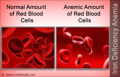
Iron deficiency anaemia (Microcytic)
Iron is a constituent of haemoglobin and rate limiting for erythropoiesis, its metabolism is dominated by its role in haemoglobin synthesis.
Iron deficiency can usually be diagnosed with the clinical history and simple laboratory tests but, a cause for the deficiency must always be sought.
Iron deficiency can be caused by long-term blood loss (e.g. gastrointestinal or uterine bleeding), Hookworm infection, poor diet, malabsorption or increased demand for iron as in pregnancy.
Clinical features
Patients with long-standing deficiency may develop nail flattening and koilonychia (concave nails), sore tongues and papillary atrophy, angular stomatitis, dysphagia and gastritis.
Patients may complain of heavy periods, indigestion or a change in bowel habit.
Iron deficiency in young children can contribute to psychomotor delay and behavioural problems.
Management
-Investigation of underlying cause.
-Correction of iron deficiency:
The normal regimen is oral ferrous sulphate 200mg three times a day for at least 6 months to replete body stores (side effects are best managed by reducing the dosage rather than changing the preparation). Parenteral iron (IV or IM) can be used where oral therapy is unsuccessful.
Megaloblastic anaemia
Megaloblastic anaemia is a common cause of a macrocytic anaemia, In clinical practice it is almost always caused by deficiency of vitamin B12 or folate.
-Vitamin B12 deficiency (Pernicious anaemia):
The classic cause is an autoimmune disorder. Patients usually have symptoms of anaemia and the generalised epithelial abnormality can manifest as glossitis and angular stomatitis.
Patients complain of unsteady gait, and if B12 deficiency is not corrected there can be a progression to irreversible damage of the central nervous system. Also, there is a possible increased incidence of carcinoma of the stomach and colorectal cancer.
Vitamin B12 levels are usually replenished by intramuscular injection of the vitamin. Several injections of 1mg hydroxycobalamin are given over the first few weeks and then either one injection every three months or daily oral vitamin B12 1 to 2mg daily for life.
Other causes of vitamin B12 deficiency are mostly abnormalities of the stomach and ileum.

-Folate deficiency:
Folate deficiency is caused by dietary insufficiency, malabsorption, excessive utilisation or a combination of these. Patients may complain of symptoms of anaemia or of an underlying disease.
Folate deficiency is treated with oral folic acid 5mg once daily, the precise duration of therapy depends on the underlying cause.
Before folate is prescribed, vitamin B12 deficiency must be excluded (or corrected) as subacute combined degeneration of the spinal cord can be precipitated.
Haemolytic anaemia
The term "haemolytic anaemia" describes a group of anaemias of differing aetiology that are all characterised by abnormal destruction of red cells.
Classification of the haemolytic anaemias:
-Hereditary spherocytosis: fluctuating levels of jaundice and palpable splenomegaly are common features. No treatment is required in patients with mild disease, in more serious cases the spleen is removed.
 -Hereditary elliptocytosis: has many similarities to hereditary spherocytosis, splenectomy helps in the rare severe cases.
2. Abnormalities of haemoglobin: thalassaemia syndromes and sickling disorders.
3. Abnormalities of red cell metabolism: glucose-6-phosphate dehydrogenase and pyruvate kinase deficiency.
-Glucose-6-phosphate (G6PD) dehydrogenase: common triggers are fava beans, drugs and infections. Treatment is to stop any offending drug and to support the patient, blood transfusion may be necessary.
-Pyruvate kinase (PK) deficiency: all general features of haemolysis can be present, but clinical symptoms are often mild for the degree of anaemia. Splenectomy may help in reducing transfusion requirements.
This disease can be divided into "warm" and "cold" types depending on the temperature at which the antibody reacts optimally with red cells.
-Warm autoimmune haemolytic anaemia (Warm AIHA): is the most common form of the disease, appoximately half of the cases are idiopathic but in the other half there is an apparent underlying cause e.g. lymphoproliferative disorders, other neoplasms, connective tissue disorders, drugs or infections.
Splenomegaly is a frequent examination finding in severe cases.
Management: identification and treatment of the cause, stop any offending drugs (e.g cephalosporin antibiotics), where the haemolysis itself requires treatment, steroids are normally used. Splenectomy is usually indicated when steroid resistance is present.
-Cold autoimmune haemolytic anaemia (Cold AIHA): the cause can be cold haemagglutinin disease, lymphoproliferative disorders, infections or paroxysmal cold haemoglobinuria.
Circularoty problems such as acrocyanosis, Raynaud´s phenomenon and ulceration may be present, haemoglobinuria may occur when red cell destruction is intravascular.
Where the Cold AIHA is chronic the mainstay of treatment is keeping the patient warm, especially in the extremities. In forms associated with lymphoproliferative disorders, cytotoxic drugs or rituximab may be helpful.

2. Isoimmune: rhesus or ABO incompatibility.
3. Non-immune and trauma: valve prostheses, microangiopathy, infection, drugs or chemicals and hypersplenism.
-Microangiopathic haemolytic anaemia (MAHA): is the intravascular destruction of red cells in the presence of an abnormal microcirculation. Common triggers are the presence of disseminated intravascular coagulation (DIC), abnormal platelet aggregation and vasculitis.
Clinical syndromes associated with MAHA include haemolytic uraemic syndrome (HUS) and thrombotic thrombocytopenic purpura (TTP).
The normal regimen is oral ferrous sulphate 200mg three times a day for at least 6 months to replete body stores (side effects are best managed by reducing the dosage rather than changing the preparation). Parenteral iron (IV or IM) can be used where oral therapy is unsuccessful.
Megaloblastic anaemia
Megaloblastic anaemia is a common cause of a macrocytic anaemia, In clinical practice it is almost always caused by deficiency of vitamin B12 or folate.
-Vitamin B12 deficiency (Pernicious anaemia):
The classic cause is an autoimmune disorder. Patients usually have symptoms of anaemia and the generalised epithelial abnormality can manifest as glossitis and angular stomatitis.
Patients complain of unsteady gait, and if B12 deficiency is not corrected there can be a progression to irreversible damage of the central nervous system. Also, there is a possible increased incidence of carcinoma of the stomach and colorectal cancer.
Vitamin B12 levels are usually replenished by intramuscular injection of the vitamin. Several injections of 1mg hydroxycobalamin are given over the first few weeks and then either one injection every three months or daily oral vitamin B12 1 to 2mg daily for life.
Other causes of vitamin B12 deficiency are mostly abnormalities of the stomach and ileum.

-Folate deficiency:
Folate deficiency is caused by dietary insufficiency, malabsorption, excessive utilisation or a combination of these. Patients may complain of symptoms of anaemia or of an underlying disease.
Folate deficiency is treated with oral folic acid 5mg once daily, the precise duration of therapy depends on the underlying cause.
Before folate is prescribed, vitamin B12 deficiency must be excluded (or corrected) as subacute combined degeneration of the spinal cord can be precipitated.
Haemolytic anaemia
The term "haemolytic anaemia" describes a group of anaemias of differing aetiology that are all characterised by abnormal destruction of red cells.
Classification of the haemolytic anaemias:
- Inherited disorders:
-Hereditary spherocytosis: fluctuating levels of jaundice and palpable splenomegaly are common features. No treatment is required in patients with mild disease, in more serious cases the spleen is removed.

2. Abnormalities of haemoglobin: thalassaemia syndromes and sickling disorders.
3. Abnormalities of red cell metabolism: glucose-6-phosphate dehydrogenase and pyruvate kinase deficiency.
-Glucose-6-phosphate (G6PD) dehydrogenase: common triggers are fava beans, drugs and infections. Treatment is to stop any offending drug and to support the patient, blood transfusion may be necessary.
-Pyruvate kinase (PK) deficiency: all general features of haemolysis can be present, but clinical symptoms are often mild for the degree of anaemia. Splenectomy may help in reducing transfusion requirements.
- Acquired disorders:
This disease can be divided into "warm" and "cold" types depending on the temperature at which the antibody reacts optimally with red cells.
-Warm autoimmune haemolytic anaemia (Warm AIHA): is the most common form of the disease, appoximately half of the cases are idiopathic but in the other half there is an apparent underlying cause e.g. lymphoproliferative disorders, other neoplasms, connective tissue disorders, drugs or infections.
Splenomegaly is a frequent examination finding in severe cases.
Management: identification and treatment of the cause, stop any offending drugs (e.g cephalosporin antibiotics), where the haemolysis itself requires treatment, steroids are normally used. Splenectomy is usually indicated when steroid resistance is present.
-Cold autoimmune haemolytic anaemia (Cold AIHA): the cause can be cold haemagglutinin disease, lymphoproliferative disorders, infections or paroxysmal cold haemoglobinuria.
Circularoty problems such as acrocyanosis, Raynaud´s phenomenon and ulceration may be present, haemoglobinuria may occur when red cell destruction is intravascular.
Where the Cold AIHA is chronic the mainstay of treatment is keeping the patient warm, especially in the extremities. In forms associated with lymphoproliferative disorders, cytotoxic drugs or rituximab may be helpful.

2. Isoimmune: rhesus or ABO incompatibility.
3. Non-immune and trauma: valve prostheses, microangiopathy, infection, drugs or chemicals and hypersplenism.
-Microangiopathic haemolytic anaemia (MAHA): is the intravascular destruction of red cells in the presence of an abnormal microcirculation. Common triggers are the presence of disseminated intravascular coagulation (DIC), abnormal platelet aggregation and vasculitis.
Clinical syndromes associated with MAHA include haemolytic uraemic syndrome (HUS) and thrombotic thrombocytopenic purpura (TTP).
Sources:
-https://themedicalpost.wordpress.com/2010/07/18/the-general-concept-of-anaemias/
-Haematology, 3rd Edition, Martin R. Howard, Churchill Livingstone, Elsevier, 2008.
-http://justinhealth.com/symptoms-of-anemia/
-http://www.spinabifida.net/cobalamin-vitamin-b12-folic-acid-deficiency-in-spina-bifida-neural-tube-defects.html
-http://www.thecambodiaherald.com/health/splenomegaly-%E2%80%93-symptoms-and-causes-688
-http://health.kernan.org/patiented/articles/000541.htm
No hay comentarios:
Publicar un comentario