martes, 29 de septiembre de 2015

ANAEMIA II

The thalassaemias
The thalassaemias are a heterogeneus group of inherited disorders of haemoglobin synthesis. They are characterised by a reduction in the rate of synthesis of either alpha or beta chains and are classified accordingly. Unbalanced synthesis of alpha and beta-globin chains can damage red cells.
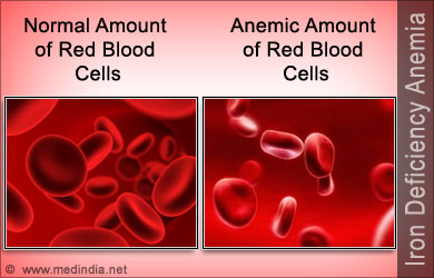
The basic haematological abnormality is a hypochromic microcytic anaemia of variable severity. The clinical severity of any thalassaemia is proportionate to the degree of imbalance of alpha and beta-globin chain synthesis.


α-Thalassaemias:


-Hb-Barts hydrops syndrome: there is a complete absence of α-chain synthesis which is needed for fetal and adult haemoglobin, the disorder is incompatible with life and death occurs in utero.

-HbH disease: there is a moderate chronic haemolytic anaemia with splenomegaly and sometimes hepatomegaly. Severe bone changes and growth retardation are unusual.

-α-Thalassaemia traits (carrier state).


β-Thalassaemias:

-β-Thalassaemia major: the characteristic severe anaemia is caused by α-chain excess leading to ineffective erythropoiesis and haemolysis. Anaemia first becomes apparent at 3-6 months, child fails to thrive and develops hepatosplenomegaly. Compensatory expansion of the marrow spaces causes atypical facies with skull bossing and maxillary enlargement. If left untreated further complications can include repeated infections, bone fractures and leg ulcers.
Blood transfusion remains the mainstay of management, splenectomy can reduce its frequency. With such regular transfusion iron chelation is necessary to minimise iron overload (accumulation of iron damages the liver, endocrine organs and heart with death), the common regimen is subcutaneous desferrioxamine given for 5-7 days per week. Compliance may be problematic but where good there is a considerably improved life expectancy.


-Thalassaemia intermedia: the clinical features are less severe than in β-Thalassaemia major. Patients usually present later (often at 2-5 years), have relatively high haemoglobin levels, moderate bone changes and normal growth. Regular transfusion is not required.

-β-Thalassaemia trait (minor): is a symptomless clinical disorder which should not be confused with iron deficiency. Genetic counselling is required in selected patients.


Sickle cell syndromes
The sickle cell syndromes are a group of haemoglobinopathies which primarily affect the Afro-Caribbean population. The common feature of these diseases is inheritance of an abnormal haemoglobin beta-chain gene, inheritance of two of these abnormal genes leads to a serious disorder termed sickle cell anaemia.


Sickle cell anaemia (HbSS): the classic form of sickle cell syndrome is enormously variable in severity.

-Haemolytic anaemia: the haemoglobin is usually in the range 60-100g/L, the symptoms of anemia are often surprisingly mild. Intercurrent infection with parvovirus or folate deficiency can cause a sudden fall in haemoglobin ("aplastic crisis").

-Vascular-occlusive crises: acute, episodic, painful crisis are a potentially disabling feature of sickle cell anemia. They may be triggered by infection or cold. Patients complain of musculoskeletal pain, hips, shoulders and vertebrae are most affected. Attacks are generally self-limiting but infarction of bone can occur. Other organs are vulnerable to infarction; most serious neurological damage which may manifest as seizures, transient ischaemic attacks and strokes. Vaso-occlusion in infancy is responsible for the "hand-foot syndrome", a type of dactylitis damaging the small bones of hand and feet.


First line treatment is rest, increased fluids and adequate oral analgesia. Constitutional upset or pain not relieved by oral analgesia requires hospital admission with continued rest, warmth, intravenous fluids and opiate analgesia.

-Sequestration crises: these arise from sickling and infarction within particular organs. Specific syndromes include "acute chest syndrome" with occlusion of the pulmonary vasculature, "girdle sequestration" caused by occlusion of the mesenteric blood supply, and hepatic and splenic sequestration.

-other complications: papillary necrosis with haematuria, loss of ability to concentrate urine, nephrotic syndrome, priapism, lower limb ulceration, proliferative retinopathy, glaucoma, liver damage and pigment gallstones.

Management:
Prophylaxis is important, patients should avoid factors known to precipitate crisis, take folate supplements and be prescribed penicillin and pneumococcal vaccine. Infections require prompt treatment.
Treatment options are blood transfusions, hydroxycarbamide and stem cell transplantation when indicated.

Prognosis:
The risk of early death is inversely related to fetal haemoglobin levels. The most common causes of death are infection in infancy, cerebro-vascular accidents in adolescence and respiratory complications in adult life.

Sickle cell trait (HbAS): normally causes no clinical problems. However, haematuria occasionally occurs as a result of renal papillary necrosis and additional care is required during pregnancy and anaesthesia.


Sources:
-http://www.slideshare.net/giridharkv/thalassemia-drkvgiridhar
-https://www.pinterest.com/kedillard/thalassemia-awareness/
-Haematology, 3rd Edition, Martin R. Howard, Churchill Livingstone, Elsevier, 2008.
-https://www.linkedin.com/pulse/sickle-cell-disease-scd-metrohealth-hmo
-http://www.slideshare.net/sandipgupta77770194/sickle-cell-disease-sandip

jueves, 24 de septiembre de 2015

ANAEMIA I

The term "anaemia" refers to haemoglobin or red cell concentration below the accepted normal range in the blood. With the widespread introduction of automated equipment into haematology laboratories the haemoglobin concentration has replaced the haematocrit as the key measurement.
The normal range for haemoglobin is affected by sex, age, ethnic group and altitude.

Normal haemoglobin (Hb) value:
-male (adult): 13 - 18 g/dL
-female (adult): 11.5 - 16 g/dL


Iron deficiency anaemia (Microcytic)
Iron is a constituent of haemoglobin and rate limiting for erythropoiesis, its metabolism is dominated by its role in haemoglobin synthesis.
Iron deficiency can usually be diagnosed with the clinical history and simple laboratory tests but, a cause for the deficiency must always be sought.

Iron deficiency can be caused by long-term blood loss (e.g. gastrointestinal or uterine bleeding), Hookworm infection, poor diet, malabsorption or increased demand for iron as in pregnancy.


Clinical features
Patients with long-standing deficiency may develop nail flattening and koilonychia (concave nails), sore tongues and papillary atrophy, angular stomatitis, dysphagia and gastritis. 
Patients may complain of heavy periods, indigestion or a change in bowel habit.
Iron deficiency in young children can contribute to psychomotor delay and behavioural problems.

Management
-Investigation of underlying cause.
-Correction of iron deficiency:
The normal regimen is oral ferrous sulphate 200mg three times a day for at least 6 months to replete body stores (side effects are best managed by reducing the dosage rather than changing the preparation). Parenteral iron (IV or IM) can be used where oral therapy is unsuccessful.

Megaloblastic anaemia 
Megaloblastic anaemia is a common cause of a macrocytic anaemia,  In clinical practice it is almost always caused by deficiency of vitamin B12 or folate.

-Vitamin B12 deficiency (Pernicious anaemia): 
The classic cause is an autoimmune disorder. Patients usually have symptoms of anaemia and the generalised epithelial abnormality can manifest as glossitis and angular stomatitis.
Patients complain of unsteady gait, and if B12 deficiency is not corrected there can be a progression to irreversible damage of the central nervous system. Also, there is a possible increased incidence of carcinoma of the stomach and colorectal cancer.
Vitamin B12 levels are usually replenished by intramuscular injection of the vitamin. Several injections of 1mg hydroxycobalamin are given over the first few weeks and then either one injection every three months or daily oral vitamin B12 1 to 2mg daily for life.
Other causes of vitamin B12 deficiency are mostly abnormalities of the stomach and ileum.


-Folate deficiency:
Folate deficiency is caused by dietary insufficiency, malabsorption, excessive utilisation or a combination of these. Patients may complain of symptoms of anaemia or of an underlying disease.
Folate deficiency is treated with oral folic acid 5mg once daily, the precise duration of therapy depends on the underlying cause.
Before folate is prescribed, vitamin B12 deficiency must be excluded (or corrected) as subacute combined degeneration of the spinal cord can be precipitated.

Haemolytic anaemia
The term "haemolytic anaemia" describes a group of anaemias of differing aetiology that are all characterised by abnormal destruction of red cells.

Classification of the haemolytic anaemias:

  • Inherited disorders:
1. Disorders of the red cell membrane: hereditary spherocytosis and hereditary elliptocytosis.
-Hereditary spherocytosis: fluctuating levels of jaundice and palpable splenomegaly are common features. No treatment is required in patients with mild disease, in more serious cases the spleen is removed.
-Hereditary elliptocytosis: has many similarities to hereditary spherocytosis, splenectomy helps in the rare severe cases.

2. Abnormalities of haemoglobin: thalassaemia syndromes and sickling disorders.

3. Abnormalities of red cell metabolism: glucose-6-phosphate dehydrogenase and pyruvate kinase deficiency.
-Glucose-6-phosphate (G6PD) dehydrogenase: common triggers are fava beans, drugs and infections. Treatment is to stop any offending drug and to support the patient, blood transfusion may be necessary.
-Pyruvate kinase (PK) deficiency: all general features of haemolysis can be present, but clinical symptoms are often mild for the degree of anaemia. Splenectomy may help in reducing transfusion requirements.
  • Acquired disorders:
1. Immune: warm and cold autoimmune haemolytic anaemia.
This disease can be divided into "warm" and "cold" types depending on the temperature at which the antibody reacts optimally with red cells.

-Warm autoimmune haemolytic anaemia (Warm AIHA): is the most common form of the disease, appoximately half of the cases are idiopathic but in the other half there is an apparent underlying cause e.g. lymphoproliferative disorders, other neoplasms, connective tissue disorders, drugs or infections.
Splenomegaly is a frequent examination finding in severe cases.
Management: identification and treatment of the cause, stop any offending drugs (e.g cephalosporin antibiotics), where the haemolysis itself requires treatment, steroids are normally used. Splenectomy is usually indicated when steroid resistance is present.

-Cold autoimmune haemolytic anaemia (Cold AIHA): the cause can be cold haemagglutinin disease, lymphoproliferative disorders, infections or paroxysmal cold haemoglobinuria.
Circularoty problems such as acrocyanosis, Raynaud´s phenomenon and ulceration may be present,  haemoglobinuria may occur when red cell destruction is intravascular.
Where the Cold AIHA is chronic the mainstay of treatment is keeping the patient warm, especially in the extremities. In forms associated with lymphoproliferative disorders, cytotoxic drugs or rituximab may be helpful.

2. Isoimmune: rhesus or ABO incompatibility.

3. Non-immune and trauma: valve prostheses, microangiopathy, infection, drugs or chemicals and hypersplenism.
-Microangiopathic haemolytic anaemia (MAHA): is the intravascular destruction of red cells in the presence of an abnormal microcirculation. Common triggers are the presence of disseminated intravascular coagulation (DIC), abnormal platelet aggregation and vasculitis.
Clinical syndromes associated with MAHA include haemolytic uraemic syndrome (HUS) and thrombotic thrombocytopenic purpura (TTP).


Sources:
-https://themedicalpost.wordpress.com/2010/07/18/the-general-concept-of-anaemias/
-Haematology, 3rd Edition, Martin R. Howard, Churchill Livingstone, Elsevier, 2008.
-http://justinhealth.com/symptoms-of-anemia/
-http://www.spinabifida.net/cobalamin-vitamin-b12-folic-acid-deficiency-in-spina-bifida-neural-tube-defects.html
-http://www.thecambodiaherald.com/health/splenomegaly-%E2%80%93-symptoms-and-causes-688
-http://health.kernan.org/patiented/articles/000541.htm

miércoles, 16 de septiembre de 2015

jueves, 10 de septiembre de 2015

BLOOD TESTS

The primary duty of the phlebotomist is to collect blood specimens for laboratory testing, this task is also performed by nurses and doctors.
Blood tests are performed for diagnostic purposes and monitor treatment, they are collected by several methods, including arterial puncture, capillary puncture and venipuncture.

Clinical laboratory services:
-Hematology: performs laboratory tests that identify diseases associated with blood and the blood-forming tissues.
-Coagulation: is the study of the ability of blood to form and dissolve clots.
-Chemistry: performs most laboratory tests and often has subsections such as toxicology and radioimmunoassay.
-Serology or Immunology:serology tests deal with the body´s response to the presence of bacterial, viral, fungal or parasitic diseases stimulating antigen-antibody reactions.
-Urinalysis: physical and chemical evaluation of urine specimens.
-Microbiology: analyzes body fluids and tissues for the presence of microorganisms, primarily by means of culture and sensitivity.
-Blood bank or Immunohematology: prepares blood products to be used for patient transfusions.


Venipuncture
Venipuncture is the process of collecting or "drawing" blood from a vein and the most common way to collect blood specimens for laboratory testing.
Common test status designations:
-Stat: immediately (from Latin statim).
-Med Emerg: medical emergency (replaces stat).
-Timed: collect at a specific time.
-ASAP: as soon as possible.
-Fasting: no food or drink except water for 8-12 hours prior to specimen collection.
-NBM, NPO: nil by mouth (from Latin nil per os).
-Preop: before an operation.
-Postop: after an operation.
-Routine: relating to established procedure.

Order of draw refers to the order in which tubes are collected during a multiple-tube draw or are filled from a syringe. CLSI (Clinical and Laboratory Standards Institute)* recommends the following order of draw for both ETS (evacuated tube system) collection and in filling tubes with a syringe:
1. Sterile tube (blood culture).
2. Blue-top coagulation tube.
3. Serum tube with or without clot activator, with or without gel.
4. Heparin tube with or without gel plasma separator.
5. EDTA tube.
6. Glycolytic inhibitor tube.


Capillary puncture
Capillary puncture can be an appropiate choice for adults and older children under the following circumstances:
-available veins are fragile or must be saved for other procedures such as chemotherapy,
-several unsuccessful venipunctures have been performed and the requested test can be collected by capillary puncture,
-the patient has thrombotic or clot-forming tendencies,
-the patient is apprehensive or has an intense fear of needles,
-there are no accessible veins,
-to obtain blood for POCT (Point-Of-Care Testing) procedures such as glucose monitoring.

Capillary puncture is the preferred method of obtaining blood from infants and very young children. The heel is the recommended site for collection of capillary puncture specimens on infants less than 1 year of age.
The CLSI order of draw for capillary specimens is as follows:
1. Blood gas specimens (CBGs)
2. EDTA specimens
3. Other additive specimens
4. Serum specimens.


Arterial puncture
Arterial puncture is technically difficult and potentially more painful and hazardous than venipuncture. The primary reason for arterial puncture is to obtain blood for arterial blood gas (ABG) tests, which evaluate respiratory function.
ABG evaluation is used in the diagnosis and management of respiratory disorders such as lung disease to provide information about a patient´s oxygenation, ventilation, acid-base balance and in the management of electrolyte and acid-base balance in patients with diabetes and other metabolic disorders.


*Organizations similar to CLSI but in the United Kingdom and another clinical laboratory information site:
-http://www.acb.org.uk/whatweare.aspx
-http://labtestsonline.org.uk/


Source:
-Phlebotomy Essential, 5th Edition, Ruth E. McCall, Cathee M. Tankersley, Wolters Kluwer/ Lippincott Williams & Wilkins, 2012.
-http://clsi.org/.
-http://www.healthaw.com/userfiles/image/Blood%20Collection.jpg
-http://i.ytimg.com/vi/YuFK22n-tvI/hqdefault.jpg

martes, 19 de mayo de 2015

SUDDEN COLLAPSE AND CARDIAC ARREST

Common causes of the sudden collapse:
-Cardiac: due to ventricular fibrillation or ventricular tachycardia in most cases, other causes can be severe bradycardia, asystole or pulmonary embolism.


-Respiratory: tension pneumothorax, aspiration/upper airway obstruction or massive haemoptysis.
-Gastrointestinal: catastrophic bleed (usually from oesophageal varices) or a ruptured abdominal aortic aneurysm.
-Anaphylaxis: a characteristic setting with a recognised trigger, wheeze, oropharyngeal swelling and severe hypotension.

Diagnosing cardiac arrest
There are four common scenarios:
1. pulseless and collapsed with ventricular fibrillation or ventricular tachycardia - cardiac arrest.
2. pulseless and collapsed with a flat trace - cardiac arrest.
3. pulseless and collapsed but with an electrical rythm (pulseless electrical activity or PEA) - cardiac arrest.
4. flat trace but patient looks well - has an electrode come off?.

THE CHAIN OF SURVIVAL
Basic life support (BLS)
Basic life support describes the process of:
-the initial assessment of the collapsed patient
-the techniques to keep the airway open
-the use of expired air ventilation and chest compression - CPR.

The primary helpers continue active resuscitation. Secondary helpers may need to fetch the crash trolley, make the medical and nursing notes available, ensure laboratory results are fed back to the arrest team, put any recent X-rays up on the viewing box, see to other patients in the ward and move any patient if appropiate and handle the relatives: phone calls and direct contact.

Sequence of actions:
1. Ensure safety of rescuer and patient
2. Shout for help. Check the patient and see if he responds.
3. If the patient responds: recovery position, check his condition (ABCDE), give oxygen, secure Iv access and attach monitors. Reassess regularly and handover to the Emergency Medical Team using a standarised communication framework (RSVP: Reason, Story, Vital signs and Plan of management).

If the patient does not respond: turn him on his back and open his airway by tilting his head and lifting his chin, remove any visible obstruction.
Keeping the airway open, look, listen and feel for normal breathing:
-if patient is breathing normally: turn him into recovery position and check for continued breathing.
-if patient is not breathing normally: check for signs of circulation and signs of life.

If there are signs of circulation, start rescue breathing, check for circulation every 10 breaths. If patient starts to breathe on his own  but remains unconscious, turn him into the recovery position.
If there are no signs of circulation or signs of life start chest compression, continuing compressions and breaths in a ratio of 30:2, at a rate of about 100-120 times a minute.
Continue resuscitation until the cardiac arrest team arrives to assist. Do not stop CPR to check the patient unless he starts to regain consciousness and shows clear signs of breathing normally again.

Advanced life support (ALS)
Once the arrest team arrives the situation can be reviewed, with the first priority being to analyse the heart rythm from the ECG monitor or via the self adhesive defibrillation pads.
In a cardiac arrest, the heart rhythm falls into one of two categories:
-Shockable rhythms: ventricular fibrillation and pulseless ventriculat tachycardia.
-Non-shockable rhythms: asystole and electrical complexes, but with no palpable pulse (PEA: pulseless electrical activity). This group needs emergency drugs (adrenaline) and continuing CPR.


Legally, the most senior doctor has the responsability of saying when to stop the resuscitation procedure.


Source:
-A nurse´s survival guide to acute medical emergencies, R. Harrison and L. Daly, Elsevier 2011

martes, 12 de mayo de 2015

MULTISYSTEM FAILURE - SHOCK

SHOCK

Classical features of shock:
-A systolic blood pressure of less than 90 mmHg
-Tachycardia
-An increased respiratory rate (>20 breaths/min) and reduced oxygen saturations
-Mottled, cool and clammy skin
-Evidence of hypovolaemia: dry mouth, dry axillary skin, postural hypotension, thirst.
-Low urine output (less than 0.5 ml/kg per h)
-Confusion or agitation progressing to coma.

Cardiogenic shock
In cardiogenic shock, the heart as a pump fails to propel blood around the circulation. The common causes are:
-heart muscle damage
-severe arrhythmia
-valve disease
-output obstruction


Hypovolaemic shock
In hypovolaemic shock, there is a reduction in the circulating blood volume due to either external fluid loss (upper gastrointestinal bleeding, severe diarrhoea and vomiting, DKA) or "internal loss" (acute pancreatitis, severe paralytic ileus).


Redistributive (low-resistance) shock
Redistributive shock is due to the effect of circulating toxins disturbing the normal distribution of blood flow within the body. Some vessels, notably those in the skin, open up and provide little or no resistance to blood flow.
Other vessels shut down in a pattern in which organs such as the kidney are starved of blood. In contrast to the other forms of shock, in redistributive shock the overall resistance to blood flow is reduced: the cardiac output is therefore high, the pulse is bounding and the peripheries are warm. The patient appears flushed and warm rather than grey and clammy. Without treatment, however, the progression is the same as that in other forms of shock - to kidney failure and to increasingly severe acidosis.
The main causes are sepsis (especially from gut and renal tract infections) and anaphylaxis.

ACUTE SEVERE HYPOTENSIVE COLLAPSE
Clinically, patients with shock present to the Acute Medical Unit as a problem of acute severe hypotensive collapse. Given this clinical situation, there are five groups of conditions to consider: cardiorespiratory collapse, massive pulmonary embolus, septicaemia, hypovolaemia and anaphylaxis.

Critical nursing tasks:
-Rapid assessment of the criticaly ill: ABCDE, maintain the safety of the patient.
-Recognition of a deteriorating patient:


-Measure the respiratory rate and the oxygen saturations.
-Measure the radial and apical pulse rates - monitor the heart.
-Measure the lying blood pressure and look for any postural decrease.
-Ensure large-bore venous access.
-Obtain an immediate 12-lead ECG.
-Assess the site and severity of any pain.
-Reassure the patient about the management plan and initiate symptom relief.
-Keep the relatives on the ward.


ANAPHYLACTIC SHOCK
Anaphylaxis is the term used for a severe generalised allergic reaction. The most common causes are foods (especially nuts), insect stings, drugs (especially antibiotics, aspirin and ACE inhibitors) and latex allergy.
From 5 min to an hour after exposure, the patient develops a generalised reaction with swellling, redness and itch. In cases of oral ingestion in food allergy, the swelling usually starts in the mouth and tongue progresses to upper airway obstruction. Occasionally, the delay from exposure to reaction is as long as 6h. 
The two potentially fatal complications are: airway obstruction and ciculatory collapse.


Emergency treatment:
-Intramuscular adrenaline (epinephrine).
-Oxygen.
-Fluids.
-Bronchodilators.
-Antihistamines.
-Steroids.
-Prevention: there must be effective education in allergen avoidance and good control of any underlying asthma by optimising the use of prophylactic anti-inflammatory treatment with inhaled steroids.

ACUTE KIDNEY INJURY (AKI)
The term Acute Kidney Injury applies to situations where there is a risk of acute renal failure and also to situations where renal failure is fully established. There are three stages in the deterioration from "at risk of" to "established" renal failure. They are defined by the urinary output:
1. Risk: urine output < 0.5ml/kg/hr for > 6 hours
2. Injury: urine output < 0.5ml/kg/hr for > 12 hours
3. Failure: urine output < 0.5ml/kg/hr for > 24 hours or anuric for 12 hours.


Nursing tasks in acute kidney injury:
-Prepare to start treatment for hyperkalaemia
-Carry out basic management of pulmonary oedema
-Identify sepsis: strong clues are fever, malaise, rigors and perhaps dysuria
-Identify hypovolaemia: symptoms of thirst and dizziness, signs of postural fall in blood pressure and low CVP (Central Venous Pressure)
-Confirm that the problem is not a simple outflow obstruction
-Take a full history
-Exclude rhabdomyolysis in prolonged immobility/coma
-Check the urine
-Ensure there is a system for reviewing the abnormal urea and electrolyte results on the ward.

Source:
-A nurse´s survival guide to acute medical emergencies, R. Harrison and L. Daly, Elsevier 2011

martes, 5 de mayo de 2015

ALCOHOL AND SUBSTANCE ABUSE

ALCOHOL ABUSE
-Acute alcohol intoxication: the main problems are hypoglycaemia, respiratory depression, airway protection and, in severe cases, fitting.
-Alcohol as a component of deliberate self-harm
-Alcohol dependence as a cause of deliberate self-harm
-Acute alcohol withdrawal syndromes: characterised by tremor, sweating, apprehension, nausea and vomiting, weakness and syncope, insomnia, auditory and visual hallucinations, fitting and severe confusion.
-Alcohol-induced disease: alcoholic gastritis, acute variceal bleeding, alcoholic liver disease or pancreatitis.
-Other alcohol-associated medical conditions: unexplained vomiting, atypical chest pain, binge-associated acute atrial fibrillation, hypertension, depression, unexplained falls and confusion in the elderly.

It is important to take a good drinking history to establish the amount and pattern of drinking and the level of dependence. 
The CAGE questionnaire:
-Have you felt you should Cut down?
-Have you been Annoyed by criticism of your drinking?
-Have you felt Guilty about your drinking?
-Do you ever have an Eye-opener in the morning to steady your nerves?
Two or more positive answers to the four questions suggests alcohol dependence.

Nursing tasks in alcohol withdrawal syndrome:
-Ensure patient´s safety.
-Check observations regularly and GCS.
-Monitor GMAWS.

-Observe for signs of trauma, nausea or pain.
-Look for signs of Wernicke´s encephalopathy : acute neuro-psychiatric reaction to severe thiamine deficiency common in alcohol misuse, characterised by acute onset of one or more of the following: acute confusional states, nystagmus and/or ataxia. It can progress to Korsakoff´s psychosis.
-Administer Pabrinex and Thiamine as prescribed, Librium (Chlordiazepoxide) should be prescribed as required.
-Provide a calm and reassuring environment as these patients are often frightened or apprehensive.


SUBSTANCE ABUSE

Cocaine
Cocaine imparts its desired effects by stimulating the release of the stress hormones (noradrenaline, adrenaline and dopamine) to inapppropriately high levels. The effects are dramatic, as may be the side-effects - notably stroke, heart attack and aortic dissection.


The nature of the cardiac damage includes cocaine-related chest pain, acute coronary syndrome, acute myocardial infarction, acute arrythmias and, with prolonged use, cocaine induced heart muscle damage. The sudden changes in pulse rate and blood pressure can trigger cerebral infarction, cerebral haemorrhage and can tear major blood vessels (aortic and coronary artery dissection).
Treatment of all of these complications should follow conventional lines except that beta-blockers are contra-indicated and that high-dose benzodiazepines may be needed to reduce cocaine-induced agitation.
  
Ecstasy
Ecstasy, or MDMA, has amphetamine-like properties which provide hyperstimulation, combined with the exertion of prolonged dancing, can result in:
-dehydration and electrolyte disturbance
-hyperarousal - agitation, tachycardia, hypertension
-muscle breakdown (rhabdomyolysis) and kidney failure
-hyperthermia up to 40°C
-convulsions
-acute liver failure
-severe acidosis

Management: reassurance and IV fluids are the main measures. Hyperthermia is managed by active cooling and with IV dantrolene 1mg/kg repeated as needed (maximum 10mg/kg). Acute agitation and convulsions respond to benzodiazepines. Acidosis may need correction with sodium bicarbonate.

Heroin abuse
Intravenous drug users are often admitted acutely with multiple complex medical, social and behavioural problems. There is frequently a conflict between the perception of the patient that withdrawal symptoms and pain are not being addressed and that of the carers who view challenging behaviour and "drug seeking". The key is an emphatic approach which is non-judgmental and focuses on the key issues of communication, co-operation and confidentiality (sometimes with the need to set defined limits to behaviour).
Heroin abuse is associated with several major health-related and psychosocial problems: HIV, hepatitis B and C, superficial and deep soft tissue sepsis, venous thromboembolic disease, opiate overdose, social isolation and crime.
Methods of heroin ingestion: inhaled (chase the dragon), smoked or injected (mainlining).


Heroin overdose
Patients are usually deeply comatose, with pin-point pupils. Treatment is with IV naloxone, the specific antidote, but reversal can be dramatic, leading to acute agitation, aggression and violence.

The current recommendations for a methadone treatment program are:
-only methadone syrup, not tablets, should be prescribed
-daily prescriptions should be given
-the effective maintenance dose should not exceed 50-100mg of methadone daily
Methadone programmes are carefully supervised and managed with a system of daily supply and directly observed ingestion. Apart from using methadone syrup, effective treatment programmes monitor drug ingestion with blood and urine testing.


Source:
-A nurse´s survival guide to acute medical emergencies, R. Harrison and L. Daly, Elsevier 2011

jueves, 30 de abril de 2015

DELIBERATE SELF-HARM AND OVERDOSES

DELIBERATE SELF-HARM
Vulnerable groups at risk of suicide:
-Recent acute psychiatric illness, particularly during the first week after discharge.
-Schizophrenia, especially in patients who are poorly compliant or lost to follow up.
-History of drug or alcohol abuse.
-Any patient who says that they are feeling suicidal, especially if accompanied by feelings of hopelessness.
-Patients living in social isolation with no family support.
-Several previous episodes of deliberate self-harm.
-A single episode of deliberate self-harm carries 1 in 20 rate of suicide within 10 years.


Type of patients admitted with deliberate self-harm:
-The embarrassed and impulsive: the spur of the moment, often triggered by a row and alcohol, not necessarily a trivial amount (often paracetamol) and low risk of recurrence or future suicide.
-The serious attempt: a planned event, carried out alone and in secret, no attempt to get help, suicide note, admits to the intended outcome, even "trivial" overdoses may represent a determined attempt at suicide.
-The recurrent attender: severe personality disorder, history of  violence, criminal convictions, drug and alcohol abuse, high risk of eventual success. 
-The high-risk self-harm: older, male, isolated, unemployed/retired, alcohol dependence, poor general health, violent method used, repeated attempts and suicide note.

Why do patients deliberately harm themselves?: to gain temporary respite, as a cry for help, as a signal of distress, communicating anger/eliciting guilt/influencing others and to end their lives.

Critical nursing tasks in deliberate self-harm:
-Care of the unconscious patient: ABCDE (check observations, GCS, ECG, gain venous access, etc).
-Take appropiate blood (glucose, alcohol, paracetamol, aspirin, LFT, INR) and urine tests (opiates, cocaine, amphetamines, ecstasy, hallucinogens).
-Decontamination of the gut: gastric lavage or activated charcoal (single dose or multiple doses).
-Ensure that all relevant information is available: mental state, past psychiatric history, previous self-harm, alcohol use, assessment of suicidal intent.
-Provide a suitable and safe environment in which to recover consciousness.


SPECIFIC OVERDOSES


  • Benzodiazepines

Benzodiazepines, unless taken as part of a more toxic cocktail, depress the consciousness level, but not sufficiently to put respiration at risk unless the patient also aspirates. Management is based on maintaining respiration and keeping a secure airway. The specific antagonist IV flumazenil reverses benzodiazepine coma immediately, but the drug has potential problems: the effects of a single dose soon wear off which may trigger acute agitation and if tricyclic antidepressants may also have been taken, flumazenil is contraindicated.


  • Paracetamol

Paracetamol remains the most common cause of acute liver failure in the UK.
For most patients 12g of paracetamol or more than 150mg per kg body weight can produce severe liver damage.

Clinical picture of severe paracetamol toxicity:
-0-24 h: minimal immediate symptoms, mild nausea and vomiting within a few hours, liver function tests normal in the first 12 h, abnormal by 18 h.
-24-48 h: right upper abdominal pain with continued vomiting, liver tenderness, progressively deranged liver function tests, prolonged prothrombin time, loin pain, haematuria, proteinuria and deteriorating kidney function.
-Days 3 and 4: maximum liver damage on fourth day, jaundice and increasing confusion with grade 3/4 hepatic encephalopathy.

Immediate management:
-Establish the exact timing of the overdose (time tablets swallowed, time help arrived, could the overdose have been staggered over several hours?).
-what else was taken?
-is this a susceptible patient (eating disorder, HIV, cystic fibrosis or malnourished, chronic alcoholic, epileptic/anti-TB drugs, underweight).
-consider activated charcoal: overdose taken within an hour and methionine is not going to be used (charcoal inactivates methionine).
-take blood for measurement of. paracetamol levels.
-start N-acetylcysteine (Parvolex) treatment if indicated (patient needs to be weighed for the correct dose). Repeat LFT´s and prothrombin time at the end of the infusion (20 h).

There is a poor prognosis 24 h after the overdose, patients should be discharged only after psychosocial assessment, after completion of N-acetylcysteine and if there are no symptoms.
Clear instructions should be given to the patient who discharges themself: to return if there is abdominal pain, persistent vomiting or confusion.


  • Antidepressants

There are two types of antidepressants used in deliberate self-harm:
-the older tricyclic antisepressants: these are dangerous and cause coma, convulsions and cardiac arrhythmias. Dothiepin is the most dangerous.
-the new generation of antidepressants (SSRIs). drugs like fluoxitine can cause "serotonin syndrome" - acute confusion, fever, muscular rigidity and twitching.
Management:
-The main dangers are: respiratory depression, cardiac arrythmias and convulsions.
-Patient remains at risk for at least 12 h after admission and are prone to arrythmias if they mobilise prematurely.
-Treatment: gastric lavage or single-dose activated charcoal.


  • Carbon monoxide poisoning

Carbon monoxide is odourless, colourless and extremely dangerous. Patients suffer hypoxic damage to the heart and central nervous system.
This gas has two important sources: incomplete combustion of carboniferous domestic fuel associated with faulty installations or accidental fires and car exhaust fumes.
Clinical picture:
-Acute exposure: headache, nausea and dizziness progressing to collapse and loss of consciousness. In severe poisonng with coma, there may be skin blistering and pressure-induced muscle damage. The limbs are spastic and there may be acute heart damage, pulmonary oedema or increased intracraneal pressure.
-Subacute exposure: slower onset of nausea, vomiting, headaches and drowsiness. The clinical picture can mimic flu or gastroenteritis - a source of confusion. As with gastroenteritis, a whole household can be affected if exposed to the same source.

Assessment and management:
-ABCDE
-Give immediate high-concentration oxygen
-Consider hyperbaric oxygen, especially in cases of pregnancy, COHb > 20% or signs of neurological dysfunction
-If it is accidental domestic exposure, the local environmental health department should be involved to assess the safety of the house.


Source:
-A nurse´s survival guide to acute medical emergencies, R. Harrison and L. Daly, Elsevier 2011

miércoles, 22 de abril de 2015

THROMBOEMBOLIC DISEASE

THROMBOSIS AND THROMBOEMBOLISATION
Thrombosis refers to the process in which a mass of clot forms inside an artery or vein. It consists of a dense network of fibrin in which are trapped variable portions of red cells and platelets.
Thrombosis blocks the vessel and impairs blood flow.
Arterial thrombosis leads to ischaemic tissue damage (myocardial infarction, stroke and peripheral vascular disease), whereas in venous thrombosis the consequences are due to back pressure and local swelling (oedema).

If the clot disintegrates, parts of it can break off, producing emboli that travel onwards: in venous thrombosis through the veins to the lungs (pulmonary embolus), and in arterial thrombosis through the arteries to major organs such as the brain (cerebral embolus), limbs (peripheral embolus), kidneys (renal infarction) and intestine (acute bowel ischaemia).

Superficial thrombophlebitis
Thrombophlebitis is a painful inflammation of the superficial veins. Classically it is seen with infected venous cannulae, but it also commonly occurs in patients with varicose veins.


The clinical picture is of palpable and very tender cords of thrombosed and inflamed veins, with the overlying skin appearing reddened or bruised.
Thrombophlebitis is not in itself dangerous, but may accompany deep vein thrombosis.
Management is removing the cause and treating the symptoms.

Deep vein thrombosis (DVT)
Deep vein thrombosis is a more serious problem, because of the risk of fatal pulmonary embolus. Most DVTs start in the deep veins of the calf, termed the distal veins, where they cause local pain and swelling. One in five extend up the leg to involve the proximal veins in the thigh and pelvis (popliteal and iliofemoral thrombosis). The risk of a pulmonary embolus from a proximal vein thrombosis is very high. A significant proportion of the survivors will develop a postphlebitic syndrome in the affected leg, with chronic swelling, varicose veins, skin pigmentation, recurrent thrombosis and venous ulceration.

The management is based on preventing progression of the thrombotic process by using heparin for an immediate effect, followed by warfarin in the medium to long term. The aim of treatment is to stabilise the situation, prevent extension, reduce the chances of embolisation and lower the risk of recurrence.

Critical nursing tasks in suspected DVT
-Give the first dose of heparin immediately.
-Ensure there is adequate pain relief.
-Be vigilant for acute ischaemia.
-Care of the pressure areas.
-Reassure the patient.
-Document all current regular and intermittent medication.
-Document the target INR (usually 2.5).
-Ensure a warfarin loading schedule is followed by the prescribing doctors.

*Other causes of a swollen painful leg: cellulitis or necrotising fasciitis.

Pulmonary thromboembolism
Most pulmonary emboli arise from the proximal veins in the thigh and pelvis. Depending on their size, emboli that travel to the lung either become wedged in the main pulmonary arteries, where they block the outflow of blood from the right side of the heart, or pass onwards to become trapped in the lung peripheries.

-The former, termed massive pulmonary emboli, produce a disastrous decrease in cardiac output, leading to sudden death or acute hypotensive collapse. Massive pulmonary emboli can move or start to break up either naturally or during the course of cardiopulmonary resuscitation. Thrombolytic drugs can help to dissolve the embolus and are used in the unstable hypotensive patient. Heparin will prevent further emboli, and as most patients who survive the first embolus die from a recurrence within the first few hours, it is important to start treatment urgently. Warfarin is added for long-term prevention.

-The smaller emboli in the periphery of the lungs produce wedge-shaped areas of lung damage and give rise to overlying pleurisy. The management of smaller peripheral emboli is based on preventing further and possibly bigger emboli by giving heparin and, subsequently, warfarin.


Classic features of a pulmonary embolus:
-Pleuritic chest pain
-Breathlessness
-Respiratory rate > 20 breaths/min
-Haemoptysis

Critical nursing tasks in acute pulmonary embolus:
-ABCDE: immediate resuscitation.
-Ensure adequate oxygenation.
-Provide an adequate circulation.
-Relieve the patient´s symptoms.
-Assess the response to treatment.
-Anticipate the need for urgent heparin therapy or thrombolysis.
-Prepare the patient for further procedures.


Source:
-A nurse´s survival guide to acute medical emergencies, R. Harrison and L. Daly, Elsevier 2011

jueves, 16 de abril de 2015

DIABETIC COMPLICATIONS

ACUTE MEDICAL CONDITIONS ASSOCIATED WITH DIABETES

  • Diabetic renal disease: one-third of Type I and between one-quarter and one-half of Type II diabetic patients develop diabetic kidney disease (diabetic nephropathy).

Urinary protein loss is often the first indication of renal disease.  An early sign that diabetes is affecting the kidneys is the presence of "micro" albuminuria - a loss of 30-300 mg per 24 hours - which can be detected using a specialised screening test.
-Proteinuria indicates probable renal involvement.
-Proteinuria goes hand-in-hand with cardiovascular complications and diabetic eye disease.
-Proteinuria is strongly associated with hypertension.
-Switching off the renin-angiotensin-aldosterone system with ACE inhibitors or angiotensin II receptor blockade slows down the deterioration from microalbuminuria to advanced diabetic nephropathy.
Important tasks if diabetic renal disease is discovered: careful blood pressure control, treatment with ACE inhibitors and related drugs, tight diabetic control, correction of lipids and advice on cessation of smoking.

  • Diabetic neuropathy: diabetic nerve damage is very common - it occurs in around one-third of patients with type II diabetes. There are two types of diabetic neuropathy:
-Peripheral neuropathy: it gives problems on the feet (numbness, poor skin nutrition and impaired mobility). The patient has no warning of trauma to the feet, nerve damage and muscle imbalance lead to foot deformity and pressure damage over abnormal bony prominences. Elsewhere in the body, nerve damage can lead to neuralgia with severe, recurrent and often undiagnosed pain in the thighs and abdomen.
-Autonomic neuropathy: it leads to abnormal regulation of involuntary muscle activity, notably in the bladder (painless retention, overflow and recurrent urinary infection), in the control of the blood pressure (severe postural hypotension) and in gastric emptying (regurgitation and aspiration of the stomach contents).


  • Cardiovascular disease: the main manifestations of cardiovascular disease are angina, myocardial infarction and heart failure. 
  • Cerebrovascular disease: the vascular damage in diabetes affects both the larger cerebral vessels and the cerebral microcirculation. Diabetic cerebrovascular disease presents with typical stroke-like symptoms due to thrombosis or hypertension-related cerebral haemorrhage. It should be assumed that any change in the consciousness level of a diabetic patient has a metabolic cause until proved otherwise. 
  • Peripheral vascular disease: it presents as an emergency with ischaemic problems in the limbs due either to acute arterial occlusion or to vascular complications in the feet: ulceration, infection and gangrene.


*The DIGAMI  regimen: an example of controlling the blood sugar in acute illness. Click here.


MANAGEMENT OF ACUTE DIABETIC EMERGENCIES

DKA (Diabetic Ketoacidosis)
Causes: infection, disruption of insulin treatment, new-onset diabetes, etc.

Underlying mechanism: in DKA there is an acute shortage of insulin. As a result, the liver produces and releases excessive amounts of glucose that appear in the blood and in the urine. The heavy glucose load in the urine pulls in water and electrolytes by osmosis, leading to losses of fluid, potassium, sodium, phosphate and magnesium. Because there is shortage of insulin, sugar cannot enter the cells  and so the body starts to burn adipose tissue as an alternative energy source. The adipose tissue breaks down to free fatty acids, which are converted into ketones by the liver. Ketones are acidic substances, so when excess ketones appear in the blood they produce an acidosis.
  
Management: adequate insulin replacement and correction of fluid and electrolyte loss.

Critical nursing tasks in DKA:             
-Observations: oxygen saturation, blood sugar, blood pressure, pulse (ECG), temperature and respiratory rate.
-Glasgow Coma Score.
-Test for urinary ketones.
-Examine for signs of infection.
-Ensure that the initial infusions are correctly prescribed and administered.
-Monitor patient´s progress and address his/her physical needs.
-Provide reassurance and support, initiate plans to prevent this from happening again.


Hyperosmolar Non-Ketonic Diabetic Coma (HONK)
This relatively rare complication is seen in the elderly Type II diabetic patients, the patient presents extremely unwell, with very high blood sugars and impaired consciousness levels, but without ketoacidosis. They are usually profoundly dehydrated due to osmotic fluid loss. The history usually involves a precipitating illness.
The diagnosis is made from the clinical setting and the finding of a high blood osmolality.
Management: CVP monitoring, nasogastric intubation to prevent aspiration and judicious fluid therapy. 
As the sodium levels are usually very high, hypotonic saline (0.45%) can be used in place of the initial isotonic saline.
Because the blood is so concentrated, the patients are at great risk of thrombosis and should be anticoagulated.


Hypoglycaemia.
Symptoms: click here .
Management:
-If the patient is conscious and cooperative: 100ml Lucozade, 6 glucose tablets or four teaspoonfuls of sugar.
-If there is an impaired consciousness level: intravenous glucose or intramuscular glucagon.


Infective complications in diabetes: the acute diabetic foot.
The severily infected foot that brings the diabetic patient in as an emergency will characteristically have cellulitis spreading from an area of ulceration. By the time the patient needs admission, the ulcer will have become infected and the whole foot may be at risk.


Signs of worsening and spreading infection:
-The foot becomes increasingly painful and tender.
-The patient looks ill.
-The skin goes purple and red or black blisters appear.
-There is an increase in: pulse, temperature, blood sugars, white cell count or respiration rate.
-The infection spreads into the leg.

Management:
-control the diabetes
-treat infection
-provide adequate pain relief
-salvage the limb if there is critical ischaemia (by-pass or angioplasty)
-prevent the worsening of any pressure damage.


Source:
-A nurse´s survival guide to acute medical emergencies, R. Harrison and L. Daly, Elsevier 2011

jueves, 9 de abril de 2015

Nausea, vomiting and acute gastrointestinal events

NAUSEA AND VOMITING
Consider the underlying mechanisms and potential causes: 
-establish the history (when and how did it start, other symptoms or illnesses, pain, has it happened before, anyone else got the same symptoms?).
-is this gastrointestinal disease (any diarrhoea, abdominal pain, obstruction, is the cause acute liver damage, alcohol abuse?)
-is it drug-related? (any new medication, digoxin toxicity or opiate side-effect?)
-could this be raised intracranial pressure? (headache, malignant disease or altered consciousness level?)
-could this be metabolic? (renal failure, sodium and calcium levels, ketoacidosis?).

Nausea and vomiting in acute medical conditions:
-Migraine: commonly accompanied by vomiting. Other causes of headache like meningitis, increased intracranial pressure and subarachnoid haemorrhage are also associated with vomiting.
-Myocardial infarction: vomiting is an important feature that distinguishes the pain of a myocardial infarction from that of angina and is often accompanied by nausea and sweating.
-Sepsis: vomiting can be the only symptom of hidden infection, particularly in infections in the kidneys and lower urinary tract.
-Acute gastric dilatation: gross gastric distension leads to upper abdominal swelling associated with nausea, often accompanied by hiccups and belching. A common outcome is sudden vomiting, aspiration an cardiac arrest.


ACUTE UPPER GASTROINTESTINAL HAEMORRHAGE

Causes:
-Helicobacter pylori infection and bleeding peptic ulcers
-Acute gastritis, duodenitis and acute erosions (stress ulcers)
-Mallory-Weiss tear:
-Oesophageal varices
-Gastric and oesophageal cancer
-Dieulafoy´s erosion: erosion of a congenitally abnormal artery in the lining of the stomach, emergecy surgery is often required.

Management:
-Ensuring the safety of the patient: ABCDE.
-Investigations: endoscopy.
-Assessing the degree of bleeding: history (presence of melaena with haematemesis, clots in the vomit and recognisable blood in the stool, bleeding accompanied by syncope, postural dizziness and overwhelming thirst suggest a major bleed) and examination (pale and clammy, tachycardia of more than 100 beats/min , systolic blood pressure of less than 100mmHg and postural hypotension).
-Risk assessment with the Rockall Score:


Important nursing tasks: 
-ensure the safety of the patient, assess the likely severity of the initial blood loss and report any further loss, save any important evidence (vomited blood, fresh melaena), report any abdominal pain.
-correct the oxygen saturation,
-secure the venous access and ensure accurate fluid balance charts,
-complete baseline observations, measure the pulse, blood pressure and respiratory rate. Report any changes on vital signs which should be checked at appropiate intervals.
-identify the signs of shock (pallor, sweating, restlessness, confusion),
-take an appropiate history, ask about liver disease and clotting disorders,
-reassure the patient and attend to patient´s basic comforts, warn about the likely need for an endoscopy.

ACUTE LIVER FAILURE AND HEPATIC ENCEPHALOPATHY
Acute liver failure can be precipitated by an acute event such as viral hepatitis or by a background of chronic liver disease with portal hypertension.

Components of acute liver failure:
-impairment of the consciousness level: the combination of poor liver function and portosystemic shunting means that toxic substances are either dealt with ineffectively by the liver or bypass it altogether. The result is a toxic encephalopathy with imapirment of the consciousness level that can progress rapidly from confusion and agitation to deep coma. It is useful to assess and follow changes using the Hepatic encephalopathy scoring system:


-bleeding varices and clotting abnormalities: patients in acute liver failure are at great risk from upper gastrointestinal bleeding due to the combination of varices and impaired clotting.
-sepsis: infection can trigger or complicate acute liver failure.
-metabolic abnormalities: the main problems are hypoglycaemia and renal failure.

Critical nursing tasks in acute liver failure:
-Ensure the safety of the patient: ABCDE.
-Gain venous access.
-Carry out top-to-toe examination.
-Liaise with patient´s family.
-Ascertain the possible causes of acute liver failure: the most common are paracetamol poisoning and acute hepatitis.
-Explain the likely interventions in the first 48 h: endoscopy, radiology, transfusions, multiple infusion lines and possible transfers.
-Obtain a drug history.

Other important nursing tasks are: determine accurate fluid balance, identify sepsis, reassure the patient and manage confusion and disorientation, carry out general nursing measures.

ACUTE ABDOMINAL PAIN
Abdominal pain is a common complaint on the acute medical ward and it is a feature of several medical conditions.

Nursing tasks:
-Establish the site and pattern of the pain.
-Assess the severity of the pain.
-Look for signs of shock (tachycardia, hypotension and increased respiratory rate).
-Examine for abdominal rigidity and peritonitis: the most obvious features of acute peritonitis are localised pain on coughing and board-like abdominal rigidity, the patient does not want to move, appears shocked and is usually vomiting. Acute peritonitis may be accompanied by signs of sepsis (hypotension, rapid thready pulse and cold, mottled extremities).
-Prepare for resuscitation: the basic requirements are oxygen, IV fluids, nasogastric intubation and urinary catheterisation.

Also, it is important to look at associated clinical features, prepare the patient for further investigations, review the previous history, the urine test results and the stool chart.


ACUTE DIARRHOEA

Infective diarrhoea:


Assessment:
-History of recent foreign travel
-Ask about other possible cases
-Have there been recent doubtful meals/mass catering...?)
-History of recent antibiotic therapy
-Is this patient at special risk?: elderly, diabetic, immunosuppressed, cardiac and kidney disease.
-Is there a history of recurrent diarrhoea?: inflammatory bowel disease.

Nursing assessment: ABCDE, assess fluid loss, look for evidence of shock and diagnosis from stool cultures.

Nursing management: 
-Infection control.
-Correct fluid deficit and keep up with the loss of fluid in the stool and vomit (fluid balance and stool chart).
-Antibiotics are rarely needed unless there are signs of complications: persistent fever with a failure to improve, development of severe colitis or shock, signs of infection, severe colitis with the risk of haemorrhage, toxic dilatation and perforation or HUS (Haemolytic Uraemic Syndrome).

Clostridium Difficile diarrhoea
The indiscriminate use of antibiotics has led to a large rise in bowel infections due to clostridium difficile. The clinical picture varies from mild diarrhoea to severe colitis with fever, systemic sepsis and death.


Diagnosis relies on two fresh stool specimens from any patient with mushy or liquid stools beng sent urgently to the labwith a request for the C.difficile toxin. The result should be available on the same day. Specific treatment is with oral metronidazole or vancomycin.

The following indicate a potentially severe infection:
-Temperature > 38.5
-More than 7 episodes of diarrhoea per 24 hours
-Pulse > 100
-WCC > 15 x 109
-A 50% rise in initial creatinine level (developing renal failure)
-Distended abdomen and no diarrhoea.

C. difficile and its spores contaminate and survive on surfaces such as commodes and bedside equipment and, in a busy ward , are easily spread by staff from one patient to another.

Enteric precautions: a single room with an en-suite commode, staff wearing gloves and apron who wash their hands with soap and water before entering and leaving the room, liquid soap and disposable wipes for the patient.


Source:
-A nurse´s survival guide to acute medical emergencies, R. Harrison and L. Daly, Elsevier 2011