martes, 19 de mayo de 2015

SUDDEN COLLAPSE AND CARDIAC ARREST

Common causes of the sudden collapse:
-Cardiac: due to ventricular fibrillation or ventricular tachycardia in most cases, other causes can be severe bradycardia, asystole or pulmonary embolism.


-Respiratory: tension pneumothorax, aspiration/upper airway obstruction or massive haemoptysis.
-Gastrointestinal: catastrophic bleed (usually from oesophageal varices) or a ruptured abdominal aortic aneurysm.
-Anaphylaxis: a characteristic setting with a recognised trigger, wheeze, oropharyngeal swelling and severe hypotension.

Diagnosing cardiac arrest
There are four common scenarios:
1. pulseless and collapsed with ventricular fibrillation or ventricular tachycardia - cardiac arrest.
2. pulseless and collapsed with a flat trace - cardiac arrest.
3. pulseless and collapsed but with an electrical rythm (pulseless electrical activity or PEA) - cardiac arrest.
4. flat trace but patient looks well - has an electrode come off?.

THE CHAIN OF SURVIVAL
Basic life support (BLS)
Basic life support describes the process of:
-the initial assessment of the collapsed patient
-the techniques to keep the airway open
-the use of expired air ventilation and chest compression - CPR.

The primary helpers continue active resuscitation. Secondary helpers may need to fetch the crash trolley, make the medical and nursing notes available, ensure laboratory results are fed back to the arrest team, put any recent X-rays up on the viewing box, see to other patients in the ward and move any patient if appropiate and handle the relatives: phone calls and direct contact.

Sequence of actions:
1. Ensure safety of rescuer and patient
2. Shout for help. Check the patient and see if he responds.
3. If the patient responds: recovery position, check his condition (ABCDE), give oxygen, secure Iv access and attach monitors. Reassess regularly and handover to the Emergency Medical Team using a standarised communication framework (RSVP: Reason, Story, Vital signs and Plan of management).

If the patient does not respond: turn him on his back and open his airway by tilting his head and lifting his chin, remove any visible obstruction.
Keeping the airway open, look, listen and feel for normal breathing:
-if patient is breathing normally: turn him into recovery position and check for continued breathing.
-if patient is not breathing normally: check for signs of circulation and signs of life.

If there are signs of circulation, start rescue breathing, check for circulation every 10 breaths. If patient starts to breathe on his own  but remains unconscious, turn him into the recovery position.
If there are no signs of circulation or signs of life start chest compression, continuing compressions and breaths in a ratio of 30:2, at a rate of about 100-120 times a minute.
Continue resuscitation until the cardiac arrest team arrives to assist. Do not stop CPR to check the patient unless he starts to regain consciousness and shows clear signs of breathing normally again.

Advanced life support (ALS)
Once the arrest team arrives the situation can be reviewed, with the first priority being to analyse the heart rythm from the ECG monitor or via the self adhesive defibrillation pads.
In a cardiac arrest, the heart rhythm falls into one of two categories:
-Shockable rhythms: ventricular fibrillation and pulseless ventriculat tachycardia.
-Non-shockable rhythms: asystole and electrical complexes, but with no palpable pulse (PEA: pulseless electrical activity). This group needs emergency drugs (adrenaline) and continuing CPR.


Legally, the most senior doctor has the responsability of saying when to stop the resuscitation procedure.


Source:
-A nurse´s survival guide to acute medical emergencies, R. Harrison and L. Daly, Elsevier 2011

martes, 12 de mayo de 2015

MULTISYSTEM FAILURE - SHOCK

SHOCK

Classical features of shock:
-A systolic blood pressure of less than 90 mmHg
-Tachycardia
-An increased respiratory rate (>20 breaths/min) and reduced oxygen saturations
-Mottled, cool and clammy skin
-Evidence of hypovolaemia: dry mouth, dry axillary skin, postural hypotension, thirst.
-Low urine output (less than 0.5 ml/kg per h)
-Confusion or agitation progressing to coma.

Cardiogenic shock
In cardiogenic shock, the heart as a pump fails to propel blood around the circulation. The common causes are:
-heart muscle damage
-severe arrhythmia
-valve disease
-output obstruction


Hypovolaemic shock
In hypovolaemic shock, there is a reduction in the circulating blood volume due to either external fluid loss (upper gastrointestinal bleeding, severe diarrhoea and vomiting, DKA) or "internal loss" (acute pancreatitis, severe paralytic ileus).


Redistributive (low-resistance) shock
Redistributive shock is due to the effect of circulating toxins disturbing the normal distribution of blood flow within the body. Some vessels, notably those in the skin, open up and provide little or no resistance to blood flow.
Other vessels shut down in a pattern in which organs such as the kidney are starved of blood. In contrast to the other forms of shock, in redistributive shock the overall resistance to blood flow is reduced: the cardiac output is therefore high, the pulse is bounding and the peripheries are warm. The patient appears flushed and warm rather than grey and clammy. Without treatment, however, the progression is the same as that in other forms of shock - to kidney failure and to increasingly severe acidosis.
The main causes are sepsis (especially from gut and renal tract infections) and anaphylaxis.

ACUTE SEVERE HYPOTENSIVE COLLAPSE
Clinically, patients with shock present to the Acute Medical Unit as a problem of acute severe hypotensive collapse. Given this clinical situation, there are five groups of conditions to consider: cardiorespiratory collapse, massive pulmonary embolus, septicaemia, hypovolaemia and anaphylaxis.

Critical nursing tasks:
-Rapid assessment of the criticaly ill: ABCDE, maintain the safety of the patient.
-Recognition of a deteriorating patient:


-Measure the respiratory rate and the oxygen saturations.
-Measure the radial and apical pulse rates - monitor the heart.
-Measure the lying blood pressure and look for any postural decrease.
-Ensure large-bore venous access.
-Obtain an immediate 12-lead ECG.
-Assess the site and severity of any pain.
-Reassure the patient about the management plan and initiate symptom relief.
-Keep the relatives on the ward.


ANAPHYLACTIC SHOCK
Anaphylaxis is the term used for a severe generalised allergic reaction. The most common causes are foods (especially nuts), insect stings, drugs (especially antibiotics, aspirin and ACE inhibitors) and latex allergy.
From 5 min to an hour after exposure, the patient develops a generalised reaction with swellling, redness and itch. In cases of oral ingestion in food allergy, the swelling usually starts in the mouth and tongue progresses to upper airway obstruction. Occasionally, the delay from exposure to reaction is as long as 6h. 
The two potentially fatal complications are: airway obstruction and ciculatory collapse.


Emergency treatment:
-Intramuscular adrenaline (epinephrine).
-Oxygen.
-Fluids.
-Bronchodilators.
-Antihistamines.
-Steroids.
-Prevention: there must be effective education in allergen avoidance and good control of any underlying asthma by optimising the use of prophylactic anti-inflammatory treatment with inhaled steroids.

ACUTE KIDNEY INJURY (AKI)
The term Acute Kidney Injury applies to situations where there is a risk of acute renal failure and also to situations where renal failure is fully established. There are three stages in the deterioration from "at risk of" to "established" renal failure. They are defined by the urinary output:
1. Risk: urine output < 0.5ml/kg/hr for > 6 hours
2. Injury: urine output < 0.5ml/kg/hr for > 12 hours
3. Failure: urine output < 0.5ml/kg/hr for > 24 hours or anuric for 12 hours.


Nursing tasks in acute kidney injury:
-Prepare to start treatment for hyperkalaemia
-Carry out basic management of pulmonary oedema
-Identify sepsis: strong clues are fever, malaise, rigors and perhaps dysuria
-Identify hypovolaemia: symptoms of thirst and dizziness, signs of postural fall in blood pressure and low CVP (Central Venous Pressure)
-Confirm that the problem is not a simple outflow obstruction
-Take a full history
-Exclude rhabdomyolysis in prolonged immobility/coma
-Check the urine
-Ensure there is a system for reviewing the abnormal urea and electrolyte results on the ward.

Source:
-A nurse´s survival guide to acute medical emergencies, R. Harrison and L. Daly, Elsevier 2011

martes, 5 de mayo de 2015

ALCOHOL AND SUBSTANCE ABUSE

ALCOHOL ABUSE
-Acute alcohol intoxication: the main problems are hypoglycaemia, respiratory depression, airway protection and, in severe cases, fitting.
-Alcohol as a component of deliberate self-harm
-Alcohol dependence as a cause of deliberate self-harm
-Acute alcohol withdrawal syndromes: characterised by tremor, sweating, apprehension, nausea and vomiting, weakness and syncope, insomnia, auditory and visual hallucinations, fitting and severe confusion.
-Alcohol-induced disease: alcoholic gastritis, acute variceal bleeding, alcoholic liver disease or pancreatitis.
-Other alcohol-associated medical conditions: unexplained vomiting, atypical chest pain, binge-associated acute atrial fibrillation, hypertension, depression, unexplained falls and confusion in the elderly.

It is important to take a good drinking history to establish the amount and pattern of drinking and the level of dependence. 
The CAGE questionnaire:
-Have you felt you should Cut down?
-Have you been Annoyed by criticism of your drinking?
-Have you felt Guilty about your drinking?
-Do you ever have an Eye-opener in the morning to steady your nerves?
Two or more positive answers to the four questions suggests alcohol dependence.

Nursing tasks in alcohol withdrawal syndrome:
-Ensure patient´s safety.
-Check observations regularly and GCS.
-Monitor GMAWS.

-Observe for signs of trauma, nausea or pain.
-Look for signs of Wernicke´s encephalopathy : acute neuro-psychiatric reaction to severe thiamine deficiency common in alcohol misuse, characterised by acute onset of one or more of the following: acute confusional states, nystagmus and/or ataxia. It can progress to Korsakoff´s psychosis.
-Administer Pabrinex and Thiamine as prescribed, Librium (Chlordiazepoxide) should be prescribed as required.
-Provide a calm and reassuring environment as these patients are often frightened or apprehensive.


SUBSTANCE ABUSE

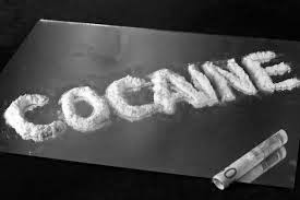
Cocaine
Cocaine imparts its desired effects by stimulating the release of the stress hormones (noradrenaline, adrenaline and dopamine) to inapppropriately high levels. The effects are dramatic, as may be the side-effects - notably stroke, heart attack and aortic dissection.


The nature of the cardiac damage includes cocaine-related chest pain, acute coronary syndrome, acute myocardial infarction, acute arrythmias and, with prolonged use, cocaine induced heart muscle damage. The sudden changes in pulse rate and blood pressure can trigger cerebral infarction, cerebral haemorrhage and can tear major blood vessels (aortic and coronary artery dissection).
Treatment of all of these complications should follow conventional lines except that beta-blockers are contra-indicated and that high-dose benzodiazepines may be needed to reduce cocaine-induced agitation.
  
Ecstasy
Ecstasy, or MDMA, has amphetamine-like properties which provide hyperstimulation, combined with the exertion of prolonged dancing, can result in:
-dehydration and electrolyte disturbance
-hyperarousal - agitation, tachycardia, hypertension
-muscle breakdown (rhabdomyolysis) and kidney failure
-hyperthermia up to 40°C
-convulsions
-acute liver failure
-severe acidosis

Management: reassurance and IV fluids are the main measures. Hyperthermia is managed by active cooling and with IV dantrolene 1mg/kg repeated as needed (maximum 10mg/kg). Acute agitation and convulsions respond to benzodiazepines. Acidosis may need correction with sodium bicarbonate.

Heroin abuse
Intravenous drug users are often admitted acutely with multiple complex medical, social and behavioural problems. There is frequently a conflict between the perception of the patient that withdrawal symptoms and pain are not being addressed and that of the carers who view challenging behaviour and "drug seeking". The key is an emphatic approach which is non-judgmental and focuses on the key issues of communication, co-operation and confidentiality (sometimes with the need to set defined limits to behaviour).
Heroin abuse is associated with several major health-related and psychosocial problems: HIV, hepatitis B and C, superficial and deep soft tissue sepsis, venous thromboembolic disease, opiate overdose, social isolation and crime.
Methods of heroin ingestion: inhaled (chase the dragon), smoked or injected (mainlining).


Heroin overdose
Patients are usually deeply comatose, with pin-point pupils. Treatment is with IV naloxone, the specific antidote, but reversal can be dramatic, leading to acute agitation, aggression and violence.

The current recommendations for a methadone treatment program are:
-only methadone syrup, not tablets, should be prescribed
-daily prescriptions should be given
-the effective maintenance dose should not exceed 50-100mg of methadone daily
Methadone programmes are carefully supervised and managed with a system of daily supply and directly observed ingestion. Apart from using methadone syrup, effective treatment programmes monitor drug ingestion with blood and urine testing.


Source:
-A nurse´s survival guide to acute medical emergencies, R. Harrison and L. Daly, Elsevier 2011