Mostrando entradas con la etiqueta RESPIRATORY SYSTEM. Mostrar todas las entradas

miércoles, 25 de marzo de 2015

RESPIRATORY MEDICINE

PRINCIPLES OF EMERGENCY TREATMENT

1. Correct the immediate and life-threatening problems:
-Hypoxia (Oxygen, ventilation).
-Acidosis (Correct high arterial carbon dioxide).
-Hypotension (fluids +- inotropic support)
2. Treat the cause:
-Pulmonary oedema (diuretics, vasodilators).
-Bronchoconstriction (bronchodilators, steroids).
-Pulmonary embolism (anticoagulants).
-Pneumonia (antibiotics).
-Tension pneumothorax (intercostal drain).
3. Prevent further attacks:
-Asthma (education).
-Pulmonary oedema (Review previous cardiac therapy).
-Pulmonary embolism (warfarin).

RESPIRATORY FAILURE
Respiratory failure describes a state in which the lungs can no longer oxygenate the blood, and is diagnosed by measuring the arterial blood gases.
Several conditions have hypoxia without carbon dioxide retention, this pattern is termed Type 1 respiratory failure.
Some examples of Type 1 respiratory failure are: the early part of an attack of asthma, the thin breathless emphysematous patient, pneumonia, pulmonary oedema (LVF) or pulmonary embolism.
With more advanced disease in which the compensatory mechanism of trying to blow off carbon dioxide has "worn out", carbon dioxide retention (hypercapnia) occurs in addition to hypoxia. This is Type II respiratory failure.
Examples of Type II respiratory failure: severe life-threatening asthma, obese oedematous patient with severe COPD, respiratory centre depression in a severe drug overdose or obesity/hypoventilation syndrome.

Principles of treatment
For the treatment of respiratory failure to be effective, it must reverse the process by correcting the cause, which, depending on the disease, may include a combination of: airway narrowing, respiratory muscle weakness, alveolar damage, respiratory infection and/or impaired respiratory effort.


ACUTE SEVERE ASTHMA
Early in an attack of asthma, a combination of necessity and fear drives breathing sufficiently hard to blow off carbon dioxide. At this point, the levels of carbon dioxide in the blood are normal or low. In contrast, oxygen uptake is impaired and there is hypoxia, even at this early stage.
Later, as breathing tires, carbon dioxide builds up, oxygen levels continue to decrease and the patient´s condition becomes critical. The increase in carbon dioxide, which can be rapid and dramatic, now causes a sudden and dangerous decrease in blood pH. If this is unchecked, a respiratory arrest will follow.

Identify patients who are at risk:
-those who are tiring (history, observation and blood gases).
-those with severe airway narrowing (peak expiratory flow rate).
-those with hypoxaemia (oxygen saturation) and a build up of carbon dioxide (blood gases).
Signs of severe asthma:
-pulse rate of more than 110 beats/min
-a respiratory rate of more than 25 breaths/min
-the patient is too breathless to complete a sentence in one breath
-a PFR between a third and a half of their best or their predicted


Critical nursing tasks during the acute attack
-Give reassurance and support, provide explanations.
-Minimise the work of breathing.
-Monitor patient´s progress - key observations and tests: pulse, respiratory rate, PFR, oxygen saturations, blood pressure, temperature, urine dipstick (for steroid induced diabetes) and sputum colour (wallpaper glue sputum is common in asthma).
-Plan to prevent this happening again.


COPD
Click here for definition.
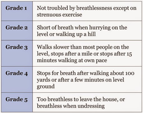
                                                         MRC breathlessness scale
Critical nursing observations:  respiratory rate, oxygen saturations, peak flow rate, pulse rate, sputum colour and temperature.
Key questions: is the patient confused? (CO2 retention), is there effective cough?, is there ankle oedema?.

Management:
-Controlled oxygen therapy.
-Bronchodilators.
-Antibiotics.
-Oral steroids.
-Non invasive ventilation.

*Normal values in blood gases:
-pH: 7.36-7.44
-pO2: 12-14 kPa
-pCO2: 4.5-6.1 kPa
-HCO3: 23-28 mmol/L


PNEUMONIA
Pneumonia is a type of repiratory infection that leads to consolidation of part of the lung. Consolidation impairs gas exchange and is visible in the chest X-ray.

Signs and symptoms-breathlessness
-cough and sputum
-cyanosis
-chest pain (usually pleurisy)
-marked tachypnoea
-fever
-systemic illness.

The severity score in pneumonia: CURB-65


Critical nursing observations: respiratory rate, blood pressure, conscioussness, oxygen saturation, pulse, temperature, sputum colour, presence of pleuritic pain, evidence of mouth sepsis or/and airways disease.
Nursing tasks: provide timely therapy, provide explanations, monitor patient´s progress, assess the need for analgesia and watch for complications.

The effect of pneumonia on the patient:
-the extent of shadowing on the chest film
-ECG, look for atrial fibrillation, a common complication
-the degree of hypoxia (pO2 less than 8 kPa)
-the degree of acidosis (pH less than 7.3)
-WCC either less than 4 or more than 20 x 10(9)/L
-blood urea more than 7 mmol/L
-low serum albumin (less than 35g/L).

Management:
-fluid balance
-oxygenation
-appropiate antibiotics
-early mobilisation and DVT prophylaxis.

Complications:
-Pleural effusion and pleural empyema.
-Aspiration pneumonia.

SPONTANEOUS PNEUMOTHORAX
A spontaneous pneumothorax occurs when a defect on the surface of the lung "pops", letting air out under positive pressure into the pleural space. This pressurised air prevents expansion of the lung and can push the mediastinal structures to the opposite side of the chest. The symptoms are pain, breathlessness and, in severe cases, cardiorespiratory collapse. 
Pneumothorax can be confused with three major conditions: pulmonary embolus, myocardial infarction and pleurisy.
A primary pneumothorax occurs in otherwise normal lungs, a secondary pneumothorax is caused by underlying lung disease or trauma.

Treatment involves removing the air from the pleural cavity by simple aspiration or, in more difficult cases, by placement of an intercostal drain.
Simple aspiration should be tried first, if aspiration does not lead to re-expansion of the lung, or if there is expansion followed by further collapse within 72hrs, then an intercostal drain will be needed.
Most of secondary pneumothoraces require tube drainage.

Nursing the patient with  a chest drain:
-Gain consent unless it is an absolute emergency,
-Prepare your patient for the sterile procedure (reassurance)  
-Positioning the patient: for example sitting in bed at 45 degrees with the appropiate arm behind his hea exposing the "triangle of safety",

-Alleviate the pain of the intercostal drain: if not contraindicated, a small dose of midazolam before skin incision can help the patient to be more relaxed and cooperative.
-Problems with intercostal drains falling out or falling apart, common problems: sutures that are too small, the weight of the connecting tubing pulling the drain out of the chest, untaped connections coming apart and failure to recognise or act on a displaced or disconnected tube.

Simple rules:
-Oxygen saturations should be monitored throughout the procedure.
-Use a transparent dressing so you can assess the state and site of the tube on at least twice a day basis.
-Bubbling tubes need to stay in and should never be clamped.
-Swinging tubes can probably be removed if the lung has expanded.
-Non-swinging tubes are often blocked.

Source:
-A nurse´s survival guide to acute medical emergencies, R. Harrison and L. Daly, Elsevier 2011.

viernes, 7 de noviembre de 2014

RESPIRATORY STIMULANTS, OXYGEN, MUCOLYTICS, COUGH PREPARATIONS AND DECONGESTANTS

RESPIRATORY STIMULANTS
Their use has been replaced by ventilatory support.
1. Doxapram hydrochloride (Dopram).

PULMONARY SURFACTANTS
Used in the management of respiratory distress syndrome in neonates and preterm neonates.
1. Beractant (Survanta).
2. Poractant Alfa (Curosurf).

OXYGEN
-High concentration oxygen therapy: for uncomplicated cases of conditions such as pneumonia, pulmonary thromboembolism, fibrosing alveolitis, shock, severe trauma, sepsis or anaphylaxis.
-Low concentration oxygen therapy: reserved for patients at risk of hypercapnic respiratory failure, which is more likely in patients with: COPD, cystic fibrosis, non-cystic fibrosis bronchiectasis, severe kyphoscoliosis or severe ankylosing apondylitis, severe lung scarring caused by tuberculosis, musculoskeletal disorders with respiratory weakness and overdose of opioids, benzodiazepines or other drugs causing respiratory depression.


MUCOLYTICS
-Carbocisteine
-Erdosteine (Erdotin)
-Mecysteine Hydrochloride (Visclair)
-Dornase Alfa (Pulmozyme)
-Hypertonic sodium chloride (MucoClear).


COUGH PREPARATIONS

1. Codeine Phosphate (Codeine Linctus).
2. Pholcodine (Pholcodine Linctus, Galenphol)
-Cough preparations used in palliative care: Methadone hydrochloride and Morphine hydrochloride.

SYSTEMIC NASAL DECONGESTANTS
-Pseudoephedrine hydrochloride (Galpseud, Sudafed).

Source:
-BNF 57 March 2009

lunes, 27 de octubre de 2014

Cortocosteroids, cromoglicate, leukotriene, antihistamines and hyposensitisation

CORTICOSTEROIDS
-Beclometasone Dipropionate (Clenil, Asmabec, Beclazone, Becodisks, Qvar, Fostair).
-Budesonide (Novolizer, Pulmicort, Symbicort).
-Ciclesonide (Alvesco).
-Fluticasone Propionate (Flixotide, Seretide).
-Mometasone Furoate (Asmanex).

CROMOGLICATE AND RELATED THERAPY AND LEUKOTRIENE RECEPTOR ANTAGONISTS

1. Cromoglicate and related therapy:
-Sodium cromoglicate (Intal).
-Nedrocromil sodium (Tilade).

2. Leukotriene receptor antagonists:
-Montelukast (Singulair).
-Zafirlukast (Accolate).

ANTIHISTAMINES, HYPOSENSITISATION AND ALLERGIC EMERGENCIES

1. Antihistamines:
-Cetirizine Hydrochloride
-Desloratadine (Neoclarityn).
-Fexofenadine Hydrochloride (Telfast).
-Levocetirizine Hydrochloride (Xyzal).
-Loratadine.
-Mizolastine (Mizollen).
-Alememazine Tartrate (Vallergan).
-Chlorphenamine Maleate (Piriton).
-Clemastine (Tavegil).
-Cyproheptadine Hydrochloride (Periactin).
-Hydroxyzine Hydrochloride (Atarax, Ucerax).
-Ketotifen (Zatiden).
-Promethazine Hydrochloride (Phenergan).

2. Allergen Inmunotherapy:
-Bee and wasp allergen extracts: Pharmaigen.
-Grass and tree pollen extracts: Pollinex, Grazax.
-Omalizumab: for prophylaxis of severe persistent allergic asthma (Xolair).

3. Allergic emergencies:


-Adrenaline (epinephrine) provides physiological reversal of the inmediate symptoms associated with hypersensitivity reactions such as anaphylaxis and angioedema (Anapen, EpiPen, Minijet Adrenaline)
-Icatibant: for acute attacks of hereditary angioedema in patients with C1 esterase inhibitor deficiency (Firazyr).


Source:
-BNF 57 March 2009

domingo, 19 de octubre de 2014

BRONCHODILATORS

ASTHMA
Asthma is a chronic disease that affects your airways. Your airways are tubes that carry air in and out of your lungs. If you have asthma, the inside walls of your airways become sore and swollen. That makes them very sensitive, and they may react strongly to things that you are allergic to or find irritating. When your airways react, they get narrower and your lungs get less air.

Symptoms of asthma include:
-Wheezing
-Coughing, especially early in the morning or at night
-Chest tightness
-Shortness of breath

Not all people who have asthma have these symptoms. Having these symptoms doesn't always mean that you have asthma. Your doctor will diagnose asthma based on lung function tests, your medical history, and a physical exam. You may also have allergy tests.
When your asthma symptoms become worse than usual, it's called an asthma attack. Severe asthma attacks may require emergency care, and they can be fatal.

COPD
COPD (chronic obstructive pulmonary disease) makes it hard for you to breathe. The two main types are chronic bronchitis and emphysema. The main cause of COPD is long-term exposure to substances that irritate and damage the lungs. This is usually cigarette smoke. Air pollution, chemical fumes, or dust can also cause it.

At first, COPD may cause no symptoms or only mild symptoms. As the disease gets worse, symptoms usually become more severe. They include:
-A cough that produces a lot of mucus
-Shortness of breath, especially with physical activity
-Wheezing
-Chest tightness

Doctors use lung function tests, imaging tests, and blood tests to diagnose COPD. There is no cure. Treatments may relieve symptoms. They include medicines, oxygen therapy, surgery, or a lung transplant. Quitting smoking is the most important step you can take to treat COPD.

CROUP
Croup is breathing difficulty and a "barking" cough. Croup is due to swelling around the vocal cords. It is common in infants and children.


PHARMACOLOGY
BRONCHODILATORS

1. Adrenoceptor agonists:
-Selective beta agonists: Bambuterol Hydrochloride (Bambec), Fenoterol Hydrobromide, Formoterol Fumarate (Atimos, Foradil, Oxis), Salbutamol (Ventolin, Ventmax, Airomir, Asmasal, Salbulin), Salmeterol (Serevent) and Terbutaline Sulphate (Bricanyl). 
-Other adrenoceptor agonists: Ephedrine Hydrochloride and Orciprenaline Sulphate (Alupent).
2. Antimuscarinic bronchodilators:
-Ipratropium Bromide (Atrovent, Respontin).
-Tiotropium (Spiriva).
3. Theophylline:
-Theophylline (Nuelin, Slo-Phyllin).
-Aminophylline (Phyllocontin Continus).
4. Compound bronchodilator preparations:
-Combivent (ipratropium bromide + salbutamol).
-Duovent (fenotrol hydrobromide + ipratropium bromide).
5. Peak flow meters, inhaler devices and nebulisers:
-Peak flow meters: standard range (MicroPeak, Mini-Wright, Personal Best, Piko-1, Pocketpeak and Vitalograph) and low range (Mini-Wright and Pocketpeak).

-Drug delivery devices: inhaler devices (pressurised metered dose inhalers, breath-actuated inhalers and dry powder inhalers) and spacer devices (Able Spacer, AeroChamber, Babyhaler, Haleraid, Nebuchamber, Nebuhaler, Optichamber, Volumatic, Pocket Chamber and PARI Vortex Spacer).
-Nebulisers: e.g: Medix Lifecare Nebuliser:


Source:
-http://www.nlm.nih.gov/medlineplus/
-BNF 57 March 2009

martes, 10 de junio de 2014

YOU ARE NOT YOUR COPD

Today, although my post was going to be about giving up smoking (in Spanish), plans have changed but not to a very different subject.
COPD is one of the long term consequences of smoking and regarding this matter, Healthline has asked me to share their initiative.
Healthline uses the power of information to make everybody healthier. They have partened with the COPD Foundation to promote a campaign called "You Are Not Your COPD" where COPD patients share their story or advice about living with the disease. For every submitted story, Heallthline will donate $10 to the COPD Foundation for research, support and treatment programs.

http://www.healthline.com/health/copd/inspirational-stories


Although, I refuse to allow any kind of advertisement in my blog, I have made an exception as I think this campaign deserves to be shared. 
Just a gesture of solidarity, good luck.

lunes, 7 de abril de 2014

COPD (Chronic Obstructive Pulmonary Disease)

COPD is a disabling condition affecting the lungs and involving irreversible lung damage, so that the lungs can no longer function at full capacity.
COPD comprises two related lung diseases:
-Chronic bronchitis: results from inflammation and irritation of the airways in the lung. This causes airway narrowing, which can cause shortness of breath or wheezing. It is characterised by the presence of cough and phlegm production for more than three months in two consecutive years.

-Emphysema; air sacs deep within the lungs, where oxygen is absorbed into the bloodstream, are prone to damage from toxins such as tobacco smoke. Emphysema develops when the air sacs enlarge and are no longer able to function properly. This results in poor oxygen delivery to the blood circulation.
The chest muscles that are involved in breathing in people with emphysema have to work harder in order to sustain an adequate oxygen level in the blood. This contributes to breathlessness, which is made worse by the associated collapse in the surrounding airways caused by the loss of the lungs natural elasticity as a consequence of lung tissue destruction. Air gets trapped in the lungs when the airways collapse during exhalation and this leads to hyperinflation of the lungs where the volume of the lungs becomes larger than normal.

There is ongoing inflammation in the airways in COPD. The onset of breathlessness is gradual over time.

Symptoms of COPD
-Many people get used to their COPD without realising it, especially in the early stages of the condition.
-You may find that you become tired easily when gardening or get out of breath when walking on an incline.
-People with COPD also tend to produce phlegm, particularly in the mornings.
-You may find that you always need to bring up phlegm by constantly coughing.
-You may also find it hard sometimes to shift phlegm from your lungs.
-The phlegm is usually clear in colour. Green or brown discoloration of thickened phlegm usually indicates an infection. Chest infection is common in people with COPD, especially during winter months. It is important to recognise infection early and to seek medical treatment.

Causes of COPD
-Smoking is the main cause of COPD.
-Other causes of COPD: alpha-1-antitrypsin deficiency, chest infections, bronchiectasis, chronic asthma and occupational exposures.

Other complications from COPD
-Breathlessness, is a frequently occuring symptom of heart failure.
-Swollen ankles, can be also a symptom of heart failure.
COPD can be very disabling; there is currently no known cure but appropriate treatment and support will help.

Treatment
The main aim of treatment is to relieve breathlessness. It does not reverse or cure the underlying condition but may improve symptoms and reduce flare-ups.
There are several forms of treatment available divided into two categories:
  • Inhaled therapy: reliever (when the need arises) and preventer (they need to be taken regularly) medication.
Class of medication:
-Corticosteroids (preventer, usually twice daily): beclometasone (AeroBec), budesonide (Pulmicort) and fluticasone (Flixotide).
-Beta-2 agonists short acting like salbutamol (Ventolin) and terbutaline (Bricanyl), they are taken when the need arises and their effects last for around 2 hours.
-Beta-2 agonists long acting like formoterol (Oxis) and salmeterol (Serevent), they keep the airways opened for up to 12 hours, used as a preventer medication usually twice a day.
-Anticholinergics short acting; can relieve breathlessness for up to 6 hours, usually used four times daily like ipratropium (Atrovent).
-Anticholinergics long acting: can keep the airway open for up to 24 hours, used once daily like tiotropium (Spiriva).

Examples of combination inhalers are:
-Combivent (ipratropium plus salbutamol).
-Seretide (fluticasone plus salmeterol).
-Symbicort (budesonide plus formoterol).

Types of inhaler devices:
-Pressurised metered-dose inhalers.

-Dry powder inhalers.

-Nebulisers.

-Using a spacer.

  • Oral therapy:
-Corticosteroids: are used mainly during a flare-up. They control inflammation of the lungs and may help in hastening recovery.

-Methylxanthines: may be used in addition to inhaler medication. They inhibit enzymes (phosphodiesterases) involved in the inflammatory process in the body and by doing so help reduce inflammation in the airways. They need to be taken daily and regular blood tests will be required. An example is theophylline (Phyllocontin).
-Mucolytics: they loosen the phlegm in the lungs making it easier to cough up. Carbocisteine (Mucodyne) and mecysteine (Visclair) are mucolytics.
-Antibiotics: may be used during a flare-up because they kill bacteria and prevent it from multiplying. They should not be used routinely to prevent infections as this may cause antibiotic resistance. Some antibiotics used are amoxicillin and clarithromycin.
-Antidepressants: may be prescribed to help with anxiety and depression related to COPD. Usually taken once daily like citalopram and paroxetine.
-Anti-phosphodiesterases: they reduce inflammation in the airways and may help with breathlessness, roflumilast and cilomilast are some examples.

  • Oxygen therapy: can be used either continuously or qhen the need arises but it may not be suitable for everyone with COPD.

Source: COPD Chronic Obstructive Pulmonary Disease, Dr Daniel Lee, Family Doctor Publications 2008.

miércoles, 1 de agosto de 2012

THE RESPIRATORY SYSTEM


Nasal cavity / Oral cavity (cavidad nasal, oral).
Top o f the throat : pharynx (faringe).
Epiglottis .
Voice box – larynx (laringe).
Windpipe –trachea (tráquea).
Bronchus, bronchi, bronchiole (bronquio, bronquios, bronquiolos).
Alveoli (alveolo).
Pleural membrane (membrane pleural).
Intercostal space (espacio intercostal).

Apnoea (apnea).
Eupnoea (eupnea).
Dyspnoea (disnea).
Bradypnoea (bradipnea).
Tachypnoea (taquipnea).

LUNG VOLUMES


ASTHMA


USING A NEBULIZER
First of all, fill the chamber of the nebulizer with the medication. Now, attach the tubing to the oxygen outlet on the wall. Next, put on the mask and tighten the elastic straps so that it fits snugly (comfortable) around the head.
After that, turn on the oxygen so the liquid medication turns into a fine mist. Finally, inhale the mist until it´s finished.


ABBREVIATIONS
@4L/min: at four litres per minute.
URTI: upper respiratory tract infection.
SOB: shortness of breath (dyspnea).
CXR: chest x-ray.
Pt ed: patient education.