miércoles, 25 de marzo de 2015

RESPIRATORY MEDICINE

PRINCIPLES OF EMERGENCY TREATMENT

1. Correct the immediate and life-threatening problems:
-Hypoxia (Oxygen, ventilation).
-Acidosis (Correct high arterial carbon dioxide).
-Hypotension (fluids +- inotropic support)
2. Treat the cause:
-Pulmonary oedema (diuretics, vasodilators).
-Bronchoconstriction (bronchodilators, steroids).
-Pulmonary embolism (anticoagulants).
-Pneumonia (antibiotics).
-Tension pneumothorax (intercostal drain).
3. Prevent further attacks:
-Asthma (education).
-Pulmonary oedema (Review previous cardiac therapy).
-Pulmonary embolism (warfarin).

RESPIRATORY FAILURE
Respiratory failure describes a state in which the lungs can no longer oxygenate the blood, and is diagnosed by measuring the arterial blood gases.
Several conditions have hypoxia without carbon dioxide retention, this pattern is termed Type 1 respiratory failure.
Some examples of Type 1 respiratory failure are: the early part of an attack of asthma, the thin breathless emphysematous patient, pneumonia, pulmonary oedema (LVF) or pulmonary embolism.
With more advanced disease in which the compensatory mechanism of trying to blow off carbon dioxide has "worn out", carbon dioxide retention (hypercapnia) occurs in addition to hypoxia. This is Type II respiratory failure.
Examples of Type II respiratory failure: severe life-threatening asthma, obese oedematous patient with severe COPD, respiratory centre depression in a severe drug overdose or obesity/hypoventilation syndrome.

Principles of treatment
For the treatment of respiratory failure to be effective, it must reverse the process by correcting the cause, which, depending on the disease, may include a combination of: airway narrowing, respiratory muscle weakness, alveolar damage, respiratory infection and/or impaired respiratory effort.


ACUTE SEVERE ASTHMA
Early in an attack of asthma, a combination of necessity and fear drives breathing sufficiently hard to blow off carbon dioxide. At this point, the levels of carbon dioxide in the blood are normal or low. In contrast, oxygen uptake is impaired and there is hypoxia, even at this early stage.
Later, as breathing tires, carbon dioxide builds up, oxygen levels continue to decrease and the patient´s condition becomes critical. The increase in carbon dioxide, which can be rapid and dramatic, now causes a sudden and dangerous decrease in blood pH. If this is unchecked, a respiratory arrest will follow.

Identify patients who are at risk:
-those who are tiring (history, observation and blood gases).
-those with severe airway narrowing (peak expiratory flow rate).
-those with hypoxaemia (oxygen saturation) and a build up of carbon dioxide (blood gases).
Signs of severe asthma:
-pulse rate of more than 110 beats/min
-a respiratory rate of more than 25 breaths/min
-the patient is too breathless to complete a sentence in one breath
-a PFR between a third and a half of their best or their predicted


Critical nursing tasks during the acute attack
-Give reassurance and support, provide explanations.
-Minimise the work of breathing.
-Monitor patient´s progress - key observations and tests: pulse, respiratory rate, PFR, oxygen saturations, blood pressure, temperature, urine dipstick (for steroid induced diabetes) and sputum colour (wallpaper glue sputum is common in asthma).
-Plan to prevent this happening again.


COPD
Click here for definition.
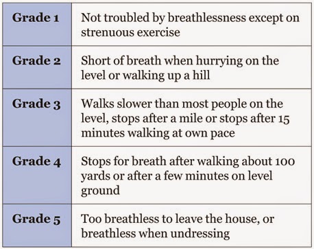
                                                         MRC breathlessness scale

Critical nursing observations:  respiratory rate, oxygen saturations, peak flow rate, pulse rate, sputum colour and temperature.
Key questions: is the patient confused? (CO2 retention), is there effective cough?, is there ankle oedema?.

Management:
-Controlled oxygen therapy.
-Bronchodilators.
-Antibiotics.
-Oral steroids.
-Non invasive ventilation.

*Normal values in blood gases:
-pH: 7.36-7.44
-pO2: 12-14 kPa
-pCO2: 4.5-6.1 kPa
-HCO3: 23-28 mmol/L


PNEUMONIA
Pneumonia is a type of repiratory infection that leads to consolidation of part of the lung. Consolidation impairs gas exchange and is visible in the chest X-ray.

Signs and symptoms-breathlessness
-cough and sputum
-cyanosis
-chest pain (usually pleurisy)
-marked tachypnoea
-fever
-systemic illness.

The severity score in pneumonia: CURB-65


Critical nursing observations: respiratory rate, blood pressure, conscioussness, oxygen saturation, pulse, temperature, sputum colour, presence of pleuritic pain, evidence of mouth sepsis or/and airways disease.
Nursing tasks: provide timely therapy, provide explanations, monitor patient´s progress, assess the need for analgesia and watch for complications.

The effect of pneumonia on the patient:
-the extent of shadowing on the chest film
-ECG, look for atrial fibrillation, a common complication
-the degree of hypoxia (pO2 less than 8 kPa)
-the degree of acidosis (pH less than 7.3)
-WCC either less than 4 or more than 20 x 10(9)/L
-blood urea more than 7 mmol/L
-low serum albumin (less than 35g/L).

Management:
-fluid balance
-oxygenation
-appropiate antibiotics
-early mobilisation and DVT prophylaxis.

Complications:
-Pleural effusion and pleural empyema.
-Aspiration pneumonia.

SPONTANEOUS PNEUMOTHORAX
A spontaneous pneumothorax occurs when a defect on the surface of the lung "pops", letting air out under positive pressure into the pleural space. This pressurised air prevents expansion of the lung and can push the mediastinal structures to the opposite side of the chest. The symptoms are pain, breathlessness and, in severe cases, cardiorespiratory collapse. 
Pneumothorax can be confused with three major conditions: pulmonary embolus, myocardial infarction and pleurisy.
A primary pneumothorax occurs in otherwise normal lungs, a secondary pneumothorax is caused by underlying lung disease or trauma.

Treatment involves removing the air from the pleural cavity by simple aspiration or, in more difficult cases, by placement of an intercostal drain.
Simple aspiration should be tried first, if aspiration does not lead to re-expansion of the lung, or if there is expansion followed by further collapse within 72hrs, then an intercostal drain will be needed.
Most of secondary pneumothoraces require tube drainage.

Nursing the patient with  a chest drain:
-Gain consent unless it is an absolute emergency,
-Prepare your patient for the sterile procedure (reassurance)  
-Positioning the patient: for example sitting in bed at 45 degrees with the appropiate arm behind his hea exposing the "triangle of safety",

-Alleviate the pain of the intercostal drain: if not contraindicated, a small dose of midazolam before skin incision can help the patient to be more relaxed and cooperative.
-Problems with intercostal drains falling out or falling apart, common problems: sutures that are too small, the weight of the connecting tubing pulling the drain out of the chest, untaped connections coming apart and failure to recognise or act on a displaced or disconnected tube.

Simple rules:
-Oxygen saturations should be monitored throughout the procedure.
-Use a transparent dressing so you can assess the state and site of the tube on at least twice a day basis.
-Bubbling tubes need to stay in and should never be clamped.
-Swinging tubes can probably be removed if the lung has expanded.
-Non-swinging tubes are often blocked.

Source:
-A nurse´s survival guide to acute medical emergencies, R. Harrison and L. Daly, Elsevier 2011.

miércoles, 18 de marzo de 2015

IMMEDIATE ASSESSMENT OF THE CRITICALLY ILL (NEWS)


  • ABCDE: IMMEDIATE ASSESSMENT AND INTERVENTION.

1. AIRWAY: check there is no problem with the airway, the airway can be maintained with a jaw thrust or chin lift. Placing the patient in the recovery position may be necessary (if no sign or suspicion of spinal injury). Use a Guedel oral airway if tolerated. Administer high-flow oxygen immediately. Look, listen and feel for expired air.

2. BREATHING: 
-Count the respiratory rate: less than 10 (imminent respiratory arrest), more than 20 (ill) and more than 30 (critically ill).
-Listen for noise and assess the effort.
-Perform oximetry.
-Continue high-flow oxygen.
-Keep the saturations greater than 90%.

3. CIRCULATION:
-Assess pulse rate and strength.
-Assess capillary refill.
-Measure the blood pressure.


4. DISABILITY:
-GCS
-Assess pupil size, equality and reaction.
-Measure blood sugar.

5. EXPOSURE:
-Look for a rash and signs of trauma.
-Check for hypothermia.
-Look in the mouth (tongue biting).


  • EARLY WARNING: TRACK AND TRIGGER SYSTEMS.
1.  MEDICAL EMERGENCY TEAM CALLING CRITERIA.


2. EARLY WARNING SCORE (EWS AND NEWS) SYSTEMS.


The NEWS system is a standarised track-and-trigger system that makes recommendations on the urgency of the clinical response required, the clinical competency of the clinical responders and the most appropiate environment for ongoing clinical care.

When a patient is acutely unwell and presents to hospital, or deteriorates and becomes acutely unwell whilst in hospital, time is of the essence and a fast and efficient clinical response is required to optomise clinical outcomes. Current evidence suggests that the triad of:
-early detection,
-timeliness of response and
-competency of the clinical response
is critical to defining clinical outcomes.

NEWS should not be used in patients under 16 years old or during pregnancy.

The National Early Warning Score system has 6 parameters:

1. Respiration rate:
-Causes of high respiration rate: acute illness and distress, generalised pain, sepsis remote from lungs, CNS disturbance and metabolic disturbances such as metabolic acidosis.
-Causes of low respiration rate: CNS depression and narcosis.

2. Oxygen saturations: assessment of pulmonary and cardiac function, it also takes into account if the patient is on any supplemental oxygen.

3. Temperature: pyrexia and hypothermia are extremes of temperature, sensitive markers of acute illness severity and physiological disturbance.

4. Systolic blood pressure: 
-Hypotension: may indicate circulatory compromise due to sepsis or volume depletion, cardiac failure or cardiac rhythm disturbance, CNS depression, hypoadrenalism and/or effect of blood pressure lowering medications. 
-Hypertension: an elevated blood pressure is an important risk factor for cardiovascular disease but it is the low systolic pressure that is most significant in the context of assessing acute illness severity.

5. Pulse rate:
-Tachycardia: may be indicative of circulatory compromise due to sepsis or volume depletion, cardiac failure, pyrexia or pain and general distress. It may be also due to cardiac arrhythmia, metabolic disturbance or drug intoxication.
-Bradycardia: may be on indicator of hypothermia, CNS depression, hypothyroidism or heart block.

6. Level of consciousness:
-A: the patient is awake.
-V: the patient responds to verbal stimulation.
-P: the patient responds to painful stimulation.
-U: the patient is completely unresponsive.

Urine output monitoring is essential for some patients as dictated by their clinical condition (e.g: sepsis) and this must be included on the fluid balance chart.

There are two mechanisms based on the NEWS which can trigger a clinical response:
-a score of 3 in any one parameter, or
-a total NEWS of 5 or more.

NEWS score: 0

NEWS score: 1-4

NEWS score: a score of 3 in any one parameter, or a total NEWS of 5 or more.

NEWS score: 7 or more.

NEWS is an aid to clinical assessment and not a substitute for competent clinical judgement. Clinical concern about a patient´s condition should always override the NEWS score.


Source:
-A nurse´s survival guide to acute medical emergencies, R. Harrison and L. Daly, Elsevier 2011.
-http://tfinews.ocbmedia.com/

martes, 10 de marzo de 2015

HEART DISEASE

The management of heart disease must be based on an understanding of the unserlying conditions, particularly ischaemic heart disease, cardiac failure and atrial fibrillation. There have been important advances in the diagnosis and management of these conditions that have altered our approach to patients:
1. Active management of myocardial infarction:
-the use of troponin-T and other markers for risk stratification.
-urgent re-perfusion by medical intervention and/or balloon angioplasty in acute myocardial infarction.
-aspirin prophylaxis.
-beta-blockers and intensive lipid-lowering therapy with statins to improve long-term outlook.
-referral of unstable patients for CABG (coronary  artery bypass grafting) or angioplasty.
2. New approaches to cardiac failure:
-the use of ACE-inhibitors.
-the use of low dose beta-blockers.
-new pacing and electrophysiological techniques.
-effective antiarrhythmic drugs: amiodarone and flecainide.
3. Recognition of the frequency and significance of atrial fibrillation:
-increased use of anticoagulation in atrial fibrillation.


HEART FAILURE

Critical nursing tasks in acute left heart failure:
-Ensure adequate oxygenation.
-Measure the blood pressure.
-Evaluate the pulse.
Important nursing tasks in acute left heart failure:
-Ensure compliance with continuous oxygen.
-Assess the response to diuretics.
-Look for symptomatic improvement.
-Demonstrate normalisation of the pulse and blood pressure.

Acute on chronic congestive cardiac failure: the main clinical features are bilateral ankle oedema, debilitating tiredness and breathlessness on exertion.
Important nursing tasks in congestive cardiac failure:
-Identify posible causes of cardiac failure (uncontrolled hypertension, ischaemic heart disease, alcohol-induced cardiac failure, chronic rheumatic valve disease)
-Establish the exact preadmission drug regimen.
-Recognise significant hypotension.
-Assess the lower limbs and pressure areas.
-Assess the fluid balance (Symptoms and signs of congestion, symptoms and signs of fluid depletion).

ISCHAEMIC HEART DISEASE
Chest pain: commonly used descriptions in angina include pressure, weight, chest tightness and constriction. Patients often also describe a feeling of breathlessness. Serious anginal episodes are accompanied by pallor, sweating and hypotension. Prolonged pain (lasting more than 15 min), especially if it is accompanied by nausea and vomiting, is more in-keeping with an infarction than with angina, although the distinction can be difficult.


Ischaemic heart disease occurs when the enlargement of cholesterol-rich plaques within the coronary artery walls lead to arterial narrowing, which is sufficient to starve the heart muscle of oxygen. Slow progressive narrowing results in stable angina. However if plaque becomes unstable and ruptures, there is a sudden combination of platelet clumping and thrombosis which clots off the coronary artery. The resulting clinical conditions are termed Acute Coronary Syndromes (ACS). There are three types of ACS dependant on the extent of the blockage and the degree of resulting heart muscle damage: STEMI, NSTEMI and unstable angina.  
Critical nursing tasks in the patient with chest pain:
-ABC triage.
-Monitor oxygen saturation.
-Obtain a 12-lead ECG.
-Take an overall look at the patient.
-Ask key questions (where id the pain? what makes it worse a what relieves it? has it occurred before?).

ATRIAL FIBRILLATION AND ARTERIAL EMBOLI
In atrial fibrillation, organised atrial activity is replaced by random electrical impulses which spread chaotically through the atrium. The atrium responds by muscular activity that can be described as a shivering motion (fibrillation), rather than a coordinated contraction. There are two important consequences:
-The atrial "squeeze", which expels the final 10% of blood into the ventricle at the end of diastole, is lost.
-The AV node is activated at random by the chaotic atrial electrical activity at rates of anything between 60 and 140 times per minute. An irregular rate of 140beats/min will not be tolerated by the heart for long.
Also, atrial fibrillation reduces cardiac output due to loss of atrial contraction, ventricular response too rapid and irregular ventricular rate.


Critical nursing tasks in atrial fibrillation:
-Assess the rate of fibrillation.
-Identify chest pain.
-Look for heart failure.
-Document hypotension.
Important nursing tasks in atrial fibrillation:
-Observe for emboli (brain, limbs, intestine).
-Look for non-cardiac triggers (pneumonia, COPD exacerbations and sepsis).
-Obtain an accurate drug history.

INFECTIVE ENDOCARDITIS
Infective endocarditis is a serious condition in which there is infection of the heart valves, usually the mitral or aortic valve. The affected valve may fail suddenly as it is destroyed by infection. Pieces of infected material can break off and spread via the blood to vital organs, resulting in:
-foci of sepsis, especially in the kidneys.
-septic cerebral emboli (stroke-like illness).

How infective endocarditis presents:
-Pyrexia of unknown origin.
-Catastrophic valve failure.
-Infected emboli.

Principles of management in the first 24h.
1. Ensure the safety of the patient.
2. Secure the diagnosis.
3. Initiate antibiotic therapy: prepare the patient for a "long haul" of antibiotic therapy (several weeks).
4. Observe for complications: allergies, left heart failure, monitor temperature and urine output and look for septic emboli.

Source:
-A nurse´s survival guide to acute medical emergencies, R. Harrison and L. Daly, Elsevier 2011.
martes, 3 de marzo de 2015

THE IMPORTANCE OF RECORD KEEPING

Principles of good record keeping:

"If it is not recorded, is not done."

1. Handwriting should be legible. 
2. All entries to records should be signed. In the case of written records, the person’s name and job title should be printed alongside the first entry. 
3. In line with local policy, you should put the date and time on all records. This should be in real time and chronological order, and be as close to the actual time as possible. 
4. Your records should be accurate and recorded in such a way that the meaning is clear
5. Records should be factual and not include unnecessary abbreviations, jargon, meaningless phrases or irrelevant speculation. 
6. You should use your professional judgement to decide what is relevant and what should be recorded. 
7. You should record details of any assessments and reviews undertaken, and provide clear evidence of the arrangements you have made for future and ongoing care. This should also include details of information given about care and treatment. 
8. Records should identify any risks or problems that have arisen and show the action taken to deal with them. 
9. You have a duty to communicate fully and effectively with your colleagues, ensuring that they have all the information they need about the people in your care. 
10. You must not alter or destroy any records without being authorised to do so. 
11. In the unlikely event that you need to alter your own or another healthcare professional’s records, you must give your name and job title, and sign and date the original documentation. You should make sure that the alterations you make, and the original record, are clear and auditable. 
12. Where appropriate, the person in your care, or their carer, should be involved in the record keeping process. 
13. The language that you use should be easily understood by the people in your care. 
14. Records should be readable when photocopied or scanned
15. You should not use coded expressions of sarcasm or humorous abbreviations to describe the people in your care. 
16. You should not falsify records.

Confidentiality.
17. You need to be fully aware of the legal requirements and guidance regarding confidentiality, and ensure your practice is in line with national and local policies. 
18.  You should be aware of the rules governing confidentiality in respect of the supply and use of data for secondary purposes. 
19. You should follow local policy and guidelines when using records for research purposes. 
20. You should not discuss the people in your care in places where you might be overheard. Nor should you leave records, either on paper or on computer screens, where they might be seen by unauthorised staff or members of the public. 
21. You should not take or keep photographs of any person, or their family, that are not clinically relevant. 

Access. 
22. People in your care should be told that information on their health records may be seen by other people or agencies involved in their care. 
23. People in your care have a right to ask to see their own health records. You should be aware of your local policy and be able to explain it to the person. 
24. People in your care have the right to ask for their information to be withheld from you or other health professionals. You must respect that right unless withholding such information would cause serious harm to that person or others. 
25. If you have any problems relating to access or record keeping, such as missing records or problems accessing records, and you cannot sort out the problem yourself, you should report the matter to someone in authority. You should keep a record that you have done so. 
26.  You should not access the records of any person, or their family, to find out personal information that is not relevant to their care. 

Disclosure. 
27. Information that can identify a person in your care must not be used or disclosed for purposes other than healthcare without the individual’s explicit consent. However, you can release this information if the law requires it, or where there is a wider public interest. 
28. Under common law, you are allowed to disclose information if it will help to prevent, detect, investigate or punish serious crime or if it will prevent abuse or serious harm to others.

Information systems. 
29. You should be aware of, and know how to use, the information systems and tools that are available to you in your practice. 
30. Smartcards or passwords to access information systems must not be shared. Similarly, do not leave systems open to access when you have finished using them. 
31. You should take reasonable measures to check that your organisation’s systems for recording and storing information, whether by computer, email, fax or any other electronic means, are secure. You should ensure you use the system appropriately, particularly in relation to confidentiality.

Personal and professional knowledge and skills. 
32. You have a duty to keep up to date with, and adhere to, relevant legislation, case law, and national and local policies relating to information and record keeping. 
33. You should be aware of, and develop, your ability to communicate effectively within teams. The way you record information and communicate is crucial. Other people will rely on your records at key communication points, especially during handover, referral and in shared care. 
34. By auditing records and acting on the results, you can assess the standard of the record keeping and communications. This will allow you to identify any areas where improvements might be made.


"In litigation, the outcome is based on proof rather than truth."
Griffith and Tengnah (2014)


Source:
-http://www.nmc-uk.org/Documents/NMC-Publications/NMC-Record-Keeping-Guidance.pdf
-Legal aspects in record keeping,  BHSCT 2015.